Hellenbrand Daniel J, Haldeman Clayton L, Lee Jae-Sung, Gableman Angela G, Dai Elena K, Ortmann Stephen D, Gotchy Jerrod C, Miller Kierra K, Doucas Adrianna M, Nowak Nicole C, Murphy William L, Hanna Amgad S
Department of Neurological Surgery; Department of Biomedical Engineering, University of Wisconsin, Madison, WI, USA.
Department of Neurological Surgery, University of Wisconsin, Madison, WI, USA.
Neural Regen Res. 2021 May;16(5):871-877. doi: 10.4103/1673-5374.297786.
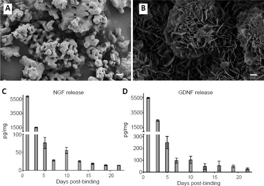
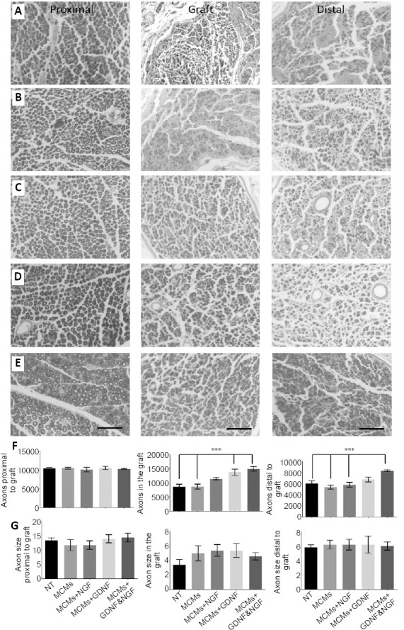
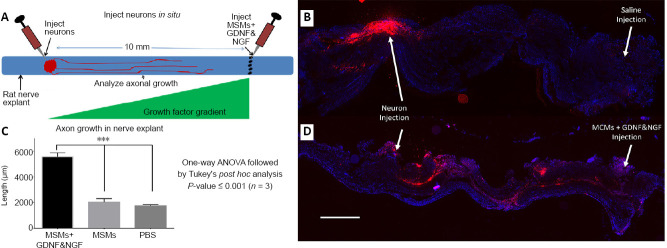
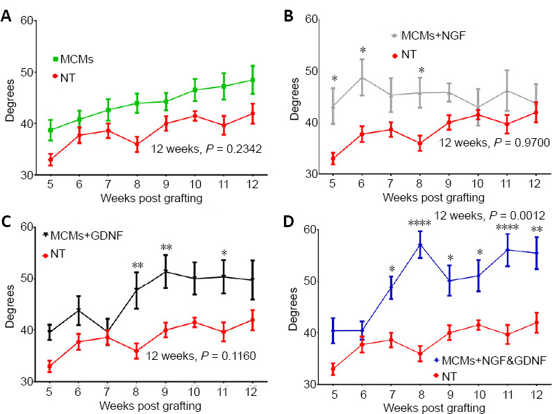
The gold standard for treating peripheral nerve injuries that have large nerve gaps where the nerves cannot be directly sutured back together because it creates tension on the nerve, is to incorporate an autologous nerve graft. However, even with the incorporation of a nerve graft, generally patients only regain a small portion of function in limbs affected by the injury. Although, there has been some promising results using growth factors to induce more axon growth through the nerve graft, many of these previous therapies are limited in their ability to release growth factors in a sustained manner and tailor them to a desired time frame. The ideal drug delivery platform would deliver growth factors at therapeutic levels for enough time to grow axons the entire length of the nerve graft. We hypothesized that mineral coated microparticles (MCMs) would bind, stabilize and release biologically active glial cell-derived neurotrophic factor (GDNF) and nerve growth factor (NGF) in a sustained manner. Therefore, the objective of this study was to test the ability of MCMs releasing growth factors at the distal end of a 10 mm sciatic nerve graft, to induce axon growth through the nerve graft and restore hind limb function. After sciatic nerve grafting in Lewis rats, the hind limb function was tested weekly by measuring the angle of the ankle at toe lift-off while walking down a track. Twelve weeks after grafting, the grafts were harvested and myelinated axons were analyzed proximal to the graft, in the center of the graft, and distal to the graft. Under physiological conditions in vitro, the MCMs delivered a burst release of NGF and GDNF for 3 days followed by a sustained release for at least 22 days. In vivo, MCMs releasing NGF and GDNF at the distal end of sciatic nerve grafts resulted in significantly more myelinated axons extending distal to the graft when compared to rats that received nerve grafts without growth factor treatment. The rats with nerve grafts incorporated with MCMs releasing NGF and GDNF also showed significant improvement in hind limb function starting at 7 weeks postoperatively and continuing through 12 weeks postoperatively when compared to rats that received nerve grafts without growth factor treatment. In conclusion, MCMs released biologically active NGF and GDNF in a sustained manner, which significantly enhanced axon growth resulting in a significant improvement of hind limb function in rats. The animal experiments were approved by University of Wisconsin-Madison Animal Care and Use Committee (ACUC, protocol# M5958) on January 3, 2018.
对于周围神经损伤且神经间隙较大、无法直接缝合以免对神经造成张力的情况,治疗的金标准是采用自体神经移植。然而,即便进行了神经移植,一般来说,患者在受损伤影响的肢体中只能恢复一小部分功能。尽管使用生长因子诱导更多轴突通过神经移植生长已取得了一些有前景的结果,但许多先前的疗法在以持续方式释放生长因子并使其适应所需时间框架的能力方面存在局限。理想的药物递送平台应以治疗水平递送生长因子足够长的时间,以使轴突在神经移植的全长范围内生长。我们假设矿物涂层微粒(MCMs)能够持续结合、稳定并释放生物活性胶质细胞源性神经营养因子(GDNF)和神经生长因子(NGF)。因此,本研究的目的是测试在10毫米坐骨神经移植远端释放生长因子的MCMs诱导轴突通过神经移植并恢复后肢功能的能力。在对Lewis大鼠进行坐骨神经移植后,每周通过测量大鼠在沿着跑道行走时脚趾离地时的踝关节角度来测试后肢功能。移植12周后,收获移植体,并对移植体近端、移植体中心以及移植体远端的有髓轴突进行分析。在体外生理条件下,MCMs在3天内实现了NGF和GDNF的爆发性释放,随后持续释放至少22天。在体内,与接受未用生长因子处理的神经移植的大鼠相比,在坐骨神经移植远端释放NGF和GDNF的MCMs导致移植体远端有更多有髓轴突延伸。与接受未用生长因子处理的神经移植的大鼠相比,植入释放NGF和GDNF的MCMs的神经移植大鼠在术后7周开始直至术后12周,后肢功能也有显著改善。总之,MCMs持续释放生物活性NGF和GDNF,显著促进了轴突生长,从而使大鼠后肢功能得到显著改善。动物实验于2018年1月3日获得威斯康星大学麦迪逊分校动物护理与使用委员会(ACUC,协议编号M5958)批准。