Pongsachareonnont Pear, Charoenphol Pulthip, Hurst Cameron, Somkijrungroj Thanapong
Vitreoretinal Research Unit, Department of Ophthalmology, Faculty of Medicine, Chulalongkorn University and King Chulalongkorn Memorial Hospital, Thai Red Cross Society, Bangkok, Thailand.
Biostatistics Center, Department of Research Affairs, Faculty of Medicine, Chulalongkorn University, Bangkok, Thailand.
Clin Ophthalmol. 2020 Nov 16;14:3871-3880. doi: 10.2147/OPTH.S270410. eCollection 2020.
This study evaluates the effect of anti-vascular endothelial growth factor (anti-VEGF) therapy on microaneurysm changes and foveal avascular zone (FAZ) using optical coherence tomography angiography (OCTA) in patients with diabetic macular edema (DME).
Prospective observational study.
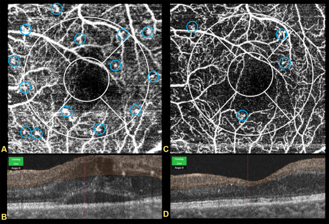
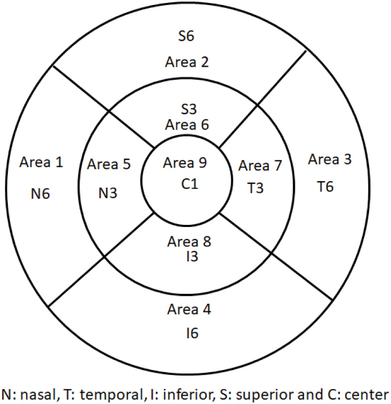
This study involved patients with DME undergoing anti-VEGF treatment (aflibercept, ranibizumab, and bevacizumab). Macula OCTA images were obtained before (visit 0) and 1 month after (visit 1) anti-VEGF injection. Microaneurysm counts were performed, and the FAZ was evaluated in the superficial capillary plexus (SCP) and deep capillary plexus (DCP). The differences in microaneurysms, FAZ, and clinical correlations were analyzed.
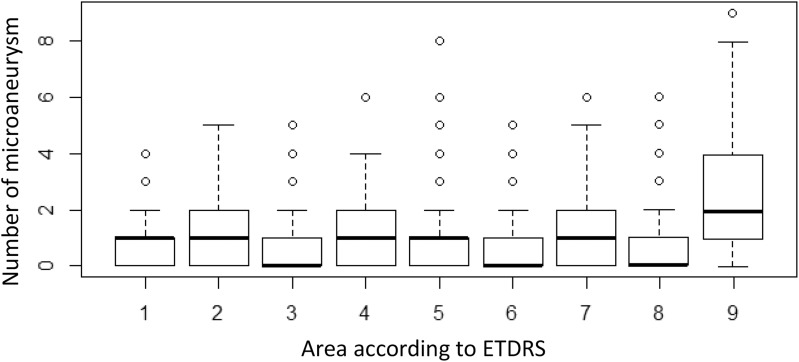
A total of 152 eyes were analyzed. The patients' mean age was 59 years. Bevacizumab was used in 69.7%, ranibizumab in 15.1%, and aflibercept in 15.1%. There was a significant reduction in the FAZ in the SCP and DCP between visits 0 and 1. All anti-VEGF medications reduced the number of microaneurysms (p<0.01). After treatment, changes in the FAZ in SCP and DCP corresponded with changes in visual acuity (p<0.01).
Microaneurysms as detected by OCTA might serve as a biomarker for a clinical response to anti-VEGF treatment in the short term. The FAZ might also predict visual acuity improvement after anti-VEGF injection.
Thai Clinical Trials Registry (TCTR20161010005).
本研究使用光学相干断层扫描血管造影(OCTA)评估抗血管内皮生长因子(抗VEGF)治疗对糖尿病性黄斑水肿(DME)患者微动脉瘤变化和黄斑无血管区(FAZ)的影响。
前瞻性观察研究。
本研究纳入接受抗VEGF治疗(阿柏西普、雷珠单抗和贝伐单抗)的DME患者。在抗VEGF注射前(就诊0)和注射后1个月(就诊1)获取黄斑OCTA图像。进行微动脉瘤计数,并在浅表毛细血管丛(SCP)和深部毛细血管丛(DCP)中评估FAZ。分析微动脉瘤、FAZ的差异及临床相关性。
共分析152只眼。患者平均年龄59岁。69.7%使用贝伐单抗,15.1%使用雷珠单抗,15.1%使用阿柏西普。就诊0和就诊1之间,SCP和DCP中的FAZ显著减小。所有抗VEGF药物均减少了微动脉瘤数量(p<0.01)。治疗后,SCP和DCP中FAZ的变化与视力变化相符(p<0.01)。
OCTA检测到的微动脉瘤可能作为抗VEGF治疗短期临床反应的生物标志物。FAZ也可能预测抗VEGF注射后视力的改善。
泰国临床试验注册中心(TCTR20161010005)。