Department of Biomedical Imaging, University Malaya Research Imaging Centre, University of Malaya, 50603, Kuala Lumpur, Malaysia.
Department of Radiology, Faculty of Medicine, University Teknologi MARA, Sungai Buloh, Selangor, Malaysia.
Sci Rep. 2020 Nov 26;10(1):20628. doi: 10.1038/s41598-020-77456-6.
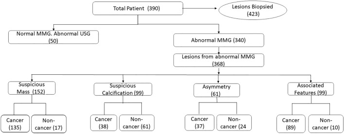
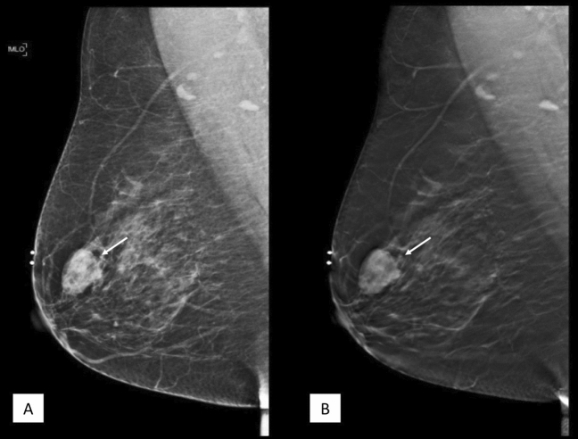
This study aims to assess the diagnostic accuracy of digital breast tomosynthesis in combination with full field digital mammography (DBT + FFDM) in the charaterisation of Breast Imaging-reporting and Data System (BI-RADS) category 3, 4 and 5 lesions. Retrospective cross-sectional study of 390 patients with BI-RADS 3, 4 and 5 mammography with available histopathology examination results were recruited from in a single center of a multi-ethnic Asian population. 2 readers independently reported the FFDM and DBT images and classified lesions detected (mass, calcifications, asymmetric density and architectural distortion) based on American College of Radiology-BI-RADS lexicon. Of the 390 patients recruited, 182 malignancies were reported. Positive predictive value (PPV) of cancer was 46.7%. The PPV in BI-RADS 4a, 4b, 4c and 5 were 6.0%, 38.3%, 68.9%, and 93.1%, respectively. Among all the cancers, 76% presented as masses, 4% as calcifications and 20% as asymmetry. An additional of 4% of cancers were detected on ultrasound. The sensitivity, specificity, PPV and NPV of mass lesions detected on DBT + FFDM were 93.8%, 85.1%, 88.8% and 91.5%, respectively. The PPV for calcification is 61.6% and asymmetry is 60.7%. 81.6% of cancer detected were invasive and 13.3% were in-situ type. Our study showed that DBT is proven to be an effective tool in the diagnosis and characterization of breast lesions and supports the current body of literature that states that integrating DBT to FFDM allows good characterization of breast lesions and accurate diagnosis of cancer.
本研究旨在评估数字乳腺断层合成摄影术(DBT)联合全数字化乳腺摄影术(FFDM)在乳腺影像报告和数据系统(BI-RADS)分类 3、4 和 5 病变中的诊断准确性。这项回顾性的病例对照研究纳入了 390 名来自单一中心的多民族亚洲人群的 BI-RADS 3、4 和 5 级乳腺摄影患者,这些患者均有可获得的组织病理学检查结果。2 位阅片者分别独立对 FFDM 和 DBT 图像进行了报告,并根据美国放射学院-BI-RADS 词汇对检测到的病变(肿块、钙化、不对称密度和结构扭曲)进行了分类。在纳入的 390 名患者中,报告了 182 例恶性肿瘤。癌症的阳性预测值(PPV)为 46.7%。BI-RADS 4a、4b、4c 和 5 的 PPV 分别为 6.0%、38.3%、68.9%和 93.1%。在所有癌症中,76%表现为肿块,4%为钙化,20%为不对称。在超声检查中还额外检出了 4%的癌症。DBT+FFDM 检测到的肿块病变的敏感性、特异性、PPV 和 NPV 分别为 93.8%、85.1%、88.8%和 91.5%。钙化的 PPV 为 61.6%,不对称的 PPV 为 60.7%。81.6%的检出癌症为浸润性癌,13.3%为原位癌。本研究表明,DBT 被证明是一种有效的诊断和评估乳腺病变的工具,支持了目前的文献,即整合 DBT 到 FFDM 可以很好地评估乳腺病变,并准确诊断癌症。