Yu Hanxi, Zhang Weichen, Han Peilin, Yang Beng, Feng Xiaode, Zhou Ping, Zhu Xiaoxu, Zhou Bingqian, Chen Wei, Qian Jianhua, Yu Jun
Department of Gynecology, The First Affiliated Hospital of Zhejiang University, School of Medicine, Hangzhou 310006, People's Republic of China.
Division of Hepatobiliary and Pancreatic Surgery, Department of Surgery First Affiliated Hospital, The First Affiliated Hospital of Zhejiang University, School of Medicine, Hangzhou 310006, People's Republic of China.
Onco Targets Ther. 2020 Nov 19;13:11935-11946. doi: 10.2147/OTT.S269168. eCollection 2020.
(), also known as serine/threonine kinase 26 (), promotes development of several cancers and is found to be highly expressed in the placenta. However, in choriocarcinoma that originated from the placenta, the expression of was undetermined and its mechanism was unknown. In this study, the expression of in choriocarcinoma as well as the underlying mechanism was explored.
To detect the expression of in patient samples and mechanism of mediating EMT by in choriocarcinoma.
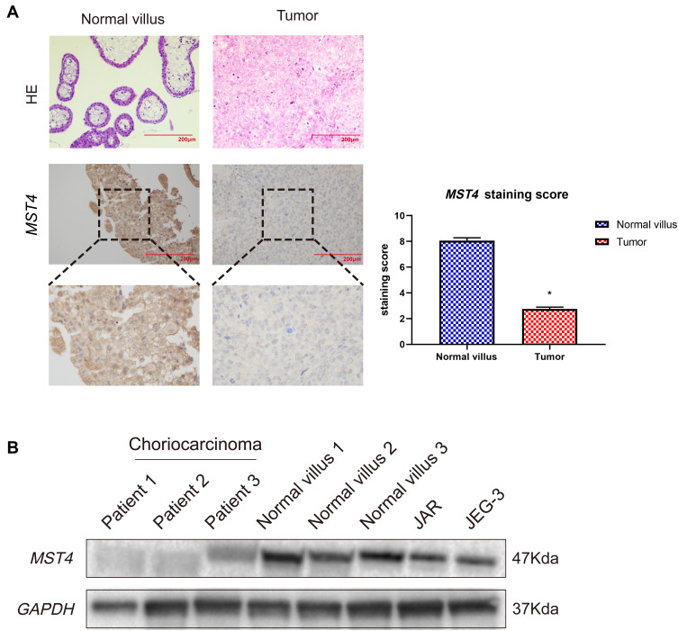
The metastatic lesions of choriocarcinoma (n=17) and volunteer villus (n=17) were collected to determine expression using immunohistochemistry and H&E staining. We use siRNA and lentiviral vector to knockdown and use plasmid to overexpress in choriocarcinoma. Then, we apply real-time polymerase chain reaction (RT-PCR), Western blot assay and immunofluorescence assay to detect target protein expressions. Cell invasion and migration and cell proliferation were detected by transwell assay and wound healing assay and CCK-8 and cell colony formation.
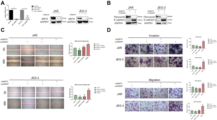
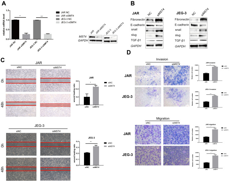
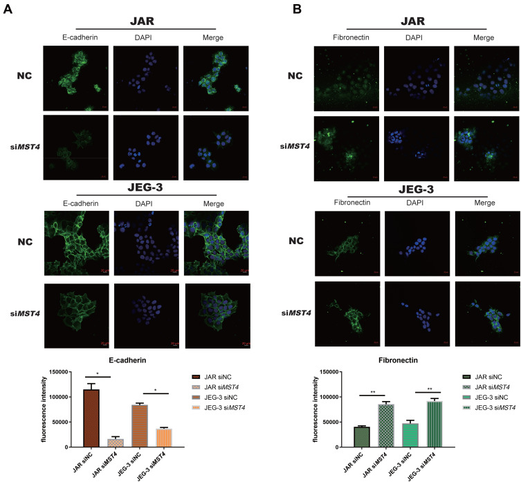
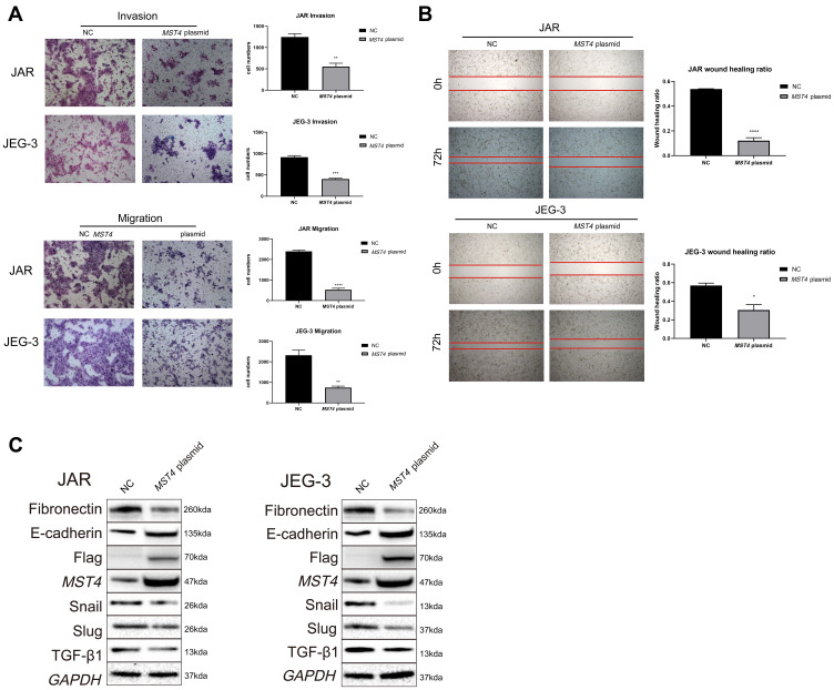
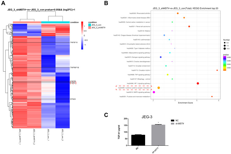
is lowly expressed in the metastatic lesions of choriocarcinoma patients when compared with normal villus. Knockdown of activated epithelial-mesenchymal transition (EMT) process, significantly increasing the ability of invasion and migration in choriocarcinoma cell lines (JAR and JEG-3). In contrast, the EMT process was restrained in choriocarcinoma cell lines with overexpressed . Meanwhile, genome-wide gene expression array, Western blot and ELISA revealed that tumor growth factor-beta 1 (TGF-β1) has significantly increased. The EMT process and metastatic prompting biofunction were reversed after using TGF-β1 inhibitor (LY364947) in the choriocarcinoma cell lines with knockdown.
Our studies demonstrated that was lowly expressed in patient samples. Additionally, JAR and JEG-3 increase cell invasion and migration ability while there is no influence on cell proliferation with knockdown. Conversely, the metastatic ability of JAR and JEG-3 was decreased with overexpressed . Moreover, TGF-β1 was a key factor after knockdown. In conclusion, affects choriocarcinoma EMT by mediating TGF-β1 expression.
(),也称为丝氨酸/苏氨酸激酶26(),促进多种癌症的发展,并且发现在胎盘中高度表达。然而,在源自胎盘的绒毛膜癌中,()的表达尚未确定,其机制也不清楚。在本研究中,探讨了()在绒毛膜癌中的表达及其潜在机制。
检测()在患者样本中的表达以及其在绒毛膜癌中介导上皮-间质转化(EMT)的机制。
收集绒毛膜癌转移灶(n = 17)和志愿者绒毛(n = 17),采用免疫组织化学和苏木精-伊红(H&E)染色确定()的表达。我们使用小干扰RNA(siRNA)和慢病毒载体敲低(),并使用质粒在绒毛膜癌中过表达()。然后,应用实时聚合酶链反应(RT-PCR)、蛋白质免疫印迹法和免疫荧光测定法检测靶蛋白表达。通过Transwell实验、伤口愈合实验以及细胞计数试剂盒-8(CCK-8)和细胞集落形成实验检测细胞侵袭、迁移和增殖情况。
与正常绒毛相比,()在绒毛膜癌患者转移灶中低表达。敲低()激活上皮-间质转化(EMT)过程,显著增加绒毛膜癌细胞系(JAR和JEG-3)的侵袭和迁移能力。相反,过表达()的绒毛膜癌细胞系中EMT过程受到抑制。同时,全基因组基因表达阵列、蛋白质免疫印迹法和酶联免疫吸附测定(ELISA)显示肿瘤生长因子-β1(TGF-β1)显著增加。在敲低()的绒毛膜癌细胞系中使用TGF-β1抑制剂(LY364947)后,EMT过程和转移促进生物功能被逆转。
我们的研究表明,()在患者样本中低表达。此外,敲低()时,JAR和JEG-3细胞的侵袭和迁移能力增加,而对细胞增殖无影响。相反,过表达()时,JAR和JEG-3的转移能力降低。此外,敲低()后,TGF-β1是关键因素。总之,()通过介导TGF-β1表达影响绒毛膜癌的EMT。