Department of Pharmacology and Therapeutics, McGill University, Montreal, QC, Canada.
Departments of Pediatrics, Ophthalmology, and Pharmacology, Hôpital Maisonneuve-Rosemont Research Center, 5415 Boul L'Assomption, Montreal, QC, H1T 2 M4, Canada.
J Neuroinflammation. 2020 Nov 27;17(1):359. doi: 10.1186/s12974-020-02032-8.
Inflammation and particularly interleukin-1β (IL-1β), a pro-inflammatory cytokine highly secreted by activated immune cells during early AMD pathological events, contribute significantly to retinal neurodegeneration. Here, we identify specific cell types that generate IL-1β and harbor the IL-1 receptor (IL-1R) and pharmacologically validate IL-1β's contribution to neuro-retinal degeneration using the IL-1R allosteric modulator composed of the amino acid sequence rytvela (as well as the orthosteric antagonist, Kineret) in a model of blue light-induced retinal degeneration.
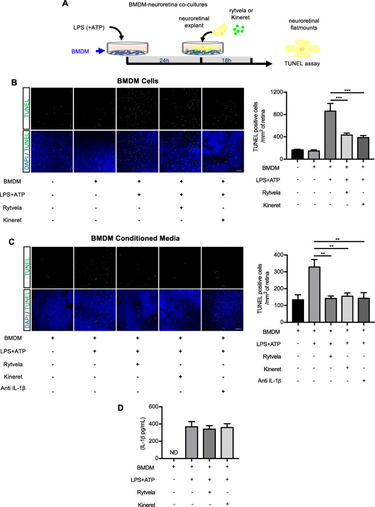
Mice were exposed to blue light for 6 h and sacrificed 3 days later. Mice were intraperitoneally injected with rytvela, Kineret, or vehicle twice daily for 3 days. The inflammatory markers F4/80, NLRP3, caspase-1, and IL-1β were assessed in the retinas. Single-cell RNA sequencing was used to determine the cell-specific expression patterns of retinal Il1b and Il1r1. Macrophage-induced photoreceptor death was assessed ex vivo using retinal explants co-cultured with LPS-activated bone marrow-derived macrophages. Photoreceptor cell death was evaluated by the TUNEL assay. Retinal function was assessed by flash electroretinography.
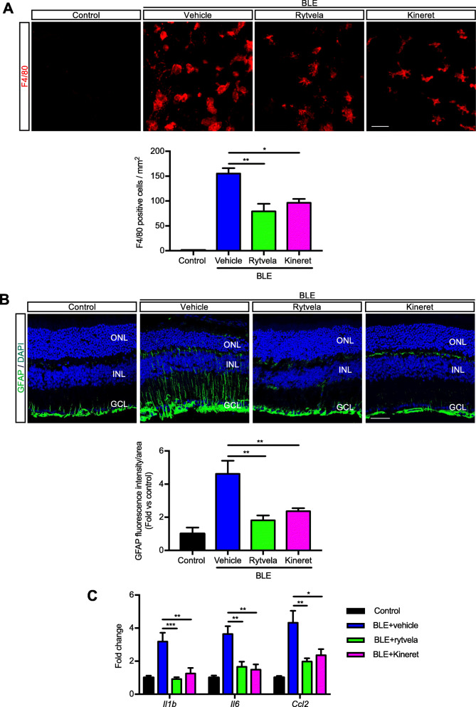
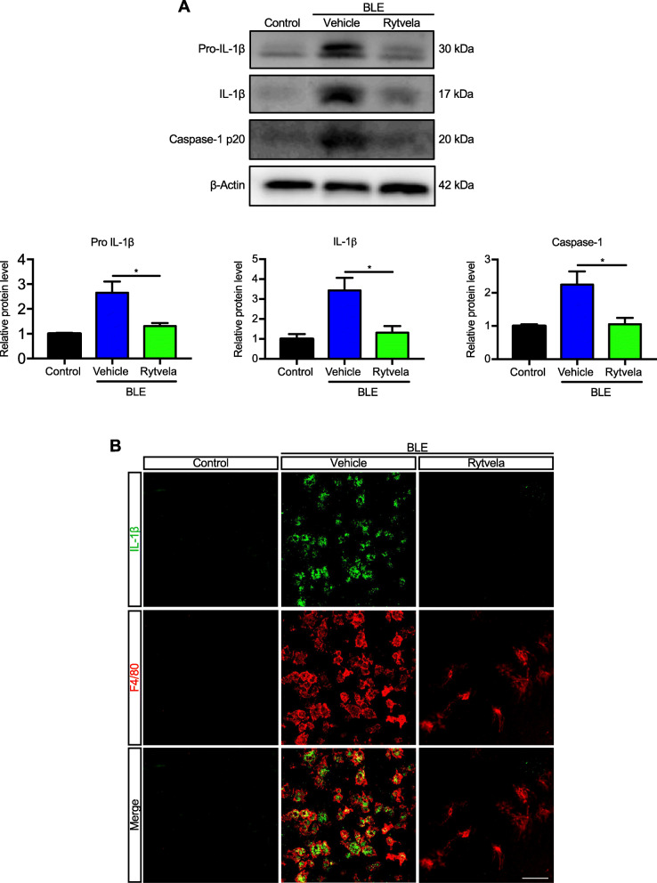
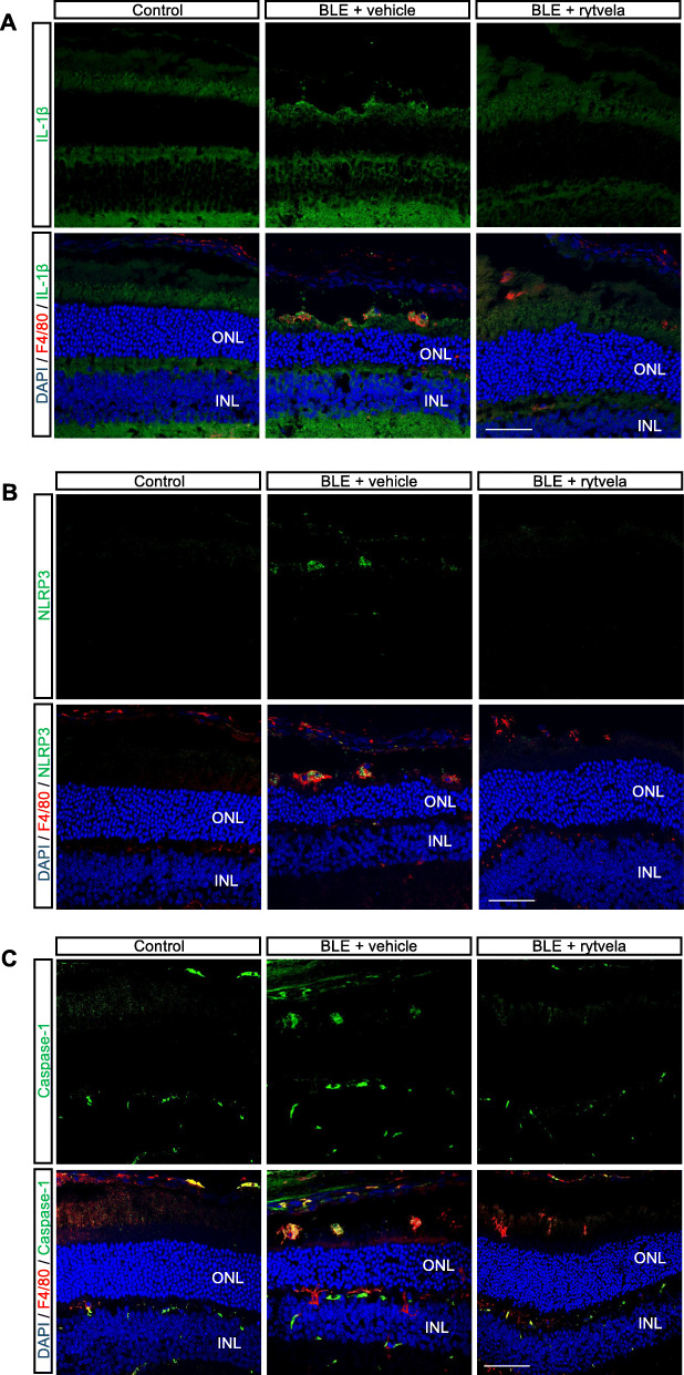
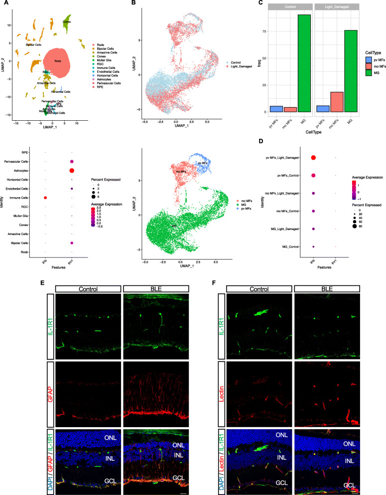
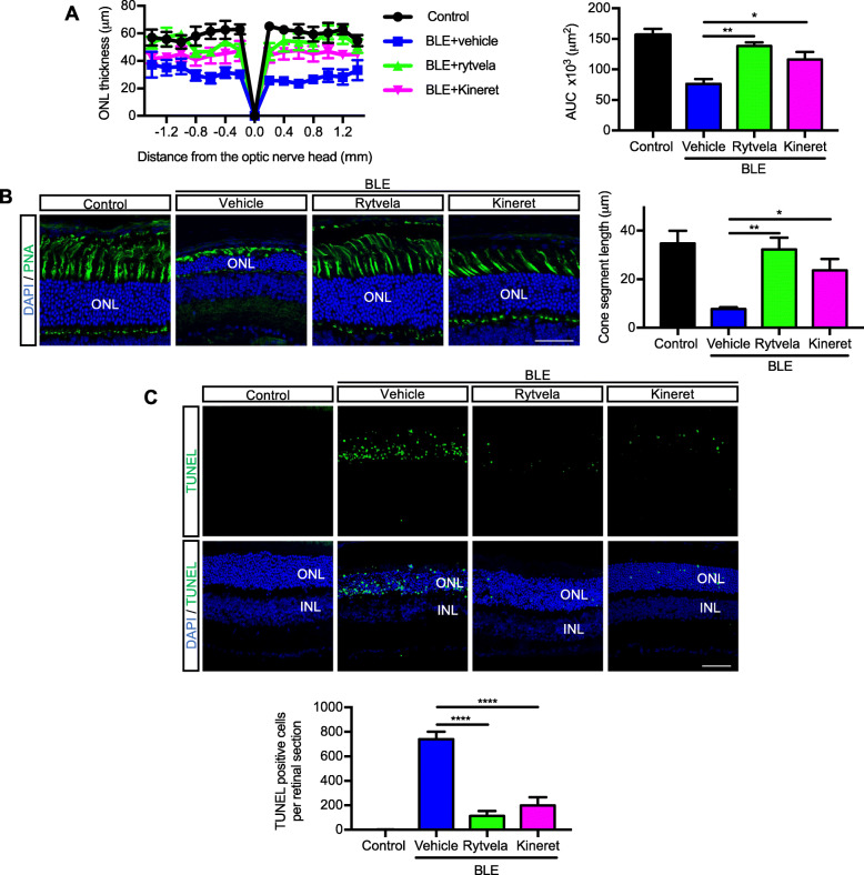
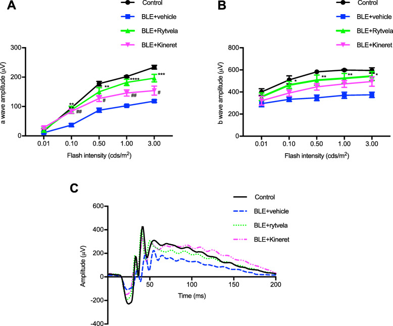
Blue light markedly increased the mononuclear phagocyte recruitment and levels of inflammatory markers associated with photoreceptor death. Co-localization of NLRP3, caspase-1, and IL-1β with F4/80 mononuclear phagocytes was clearly detected in the subretinal space, suggesting that these inflammatory cells are the main source of IL-1β. Single-cell RNA sequencing confirmed the immune-specific expression of Il1b and notably perivascular macrophages in light-challenged mice, while Il1r1 expression was found primarily in astrocytes, bipolar, and vascular cells. Retinal explants co-cultured with LPS/ATP-activated bone marrow-derived macrophages displayed a high number of TUNEL-positive photoreceptors, which was abrogated by rytvela treatment. IL-1R antagonism significantly mitigated the inflammatory response triggered in vivo by blue light exposure, and rytvela was superior to Kineret in preserving photoreceptor density and retinal function.
These findings substantiate the importance of IL-1β in neuro-retinal degeneration and revealed specific sources of Il1b from perivascular MPs, with its receptor Ilr1 being separately expressed on surrounding neuro-vascular and astroglial cells. They also validate the efficacy of rytvela-induced IL-1R modulation in suppressing detrimental inflammatory responses and preserving photoreceptor density and function in these conditions, reinforcing the rationale for clinical translation.
在 AMD 病理事件早期,激活的免疫细胞高度分泌的炎症因子,特别是白细胞介素-1β(IL-1β),对视网膜神经退行性变有重要贡献。在这里,我们确定了产生 IL-1β的特定细胞类型,并鉴定了其携带的 IL-1 受体(IL-1R),然后使用由氨基酸序列 rytvela 组成的 IL-1β 别构调节剂(以及同型拮抗剂 Kineret),在蓝光诱导的视网膜变性模型中对其对神经视网膜变性的作用进行了药理学验证。
将小鼠暴露于蓝光 6 小时,然后在 3 天后处死。将小鼠每天两次腹膜内注射 rytvela、Kineret 或载体,持续 3 天。评估视网膜中的炎症标志物 F4/80、NLRP3、半胱天冬酶-1 和 IL-1β。使用单细胞 RNA 测序确定视网膜 Il1b 和 Il1r1 的细胞特异性表达模式。使用与 LPS 激活的骨髓来源的巨噬细胞共培养的视网膜外植体评估巨噬细胞诱导的光感受器死亡。通过 TUNEL 测定评估光感受器细胞死亡。通过闪光视网膜电图评估视网膜功能。
蓝光显著增加了单核吞噬细胞的募集和与光感受器死亡相关的炎症标志物的水平。在视网膜下空间中清楚地检测到 NLRP3、半胱天冬酶-1 和 IL-1β与 F4/80 单核吞噬细胞的共定位,表明这些炎症细胞是 IL-1β 的主要来源。单细胞 RNA 测序证实,在受光挑战的小鼠中,Il1b 具有免疫特异性表达,尤其是血管周围巨噬细胞,而 Il1r1 表达主要在星形胶质细胞、双极细胞和血管细胞中。与 LPS/ATP 激活的骨髓来源的巨噬细胞共培养的视网膜外植体显示出大量 TUNEL 阳性光感受器,而 rytvela 处理则消除了这一现象。IL-1R 拮抗作用显著减轻了蓝光暴露引起的体内炎症反应,并且在保护光感受器密度和视网膜功能方面,rytvela 优于 Kineret。
这些发现证实了 IL-1β 在神经视网膜变性中的重要性,并揭示了血管周围 MPs 中 Il1b 的特定来源,其受体 Ilr1 分别在周围的神经血管和星形胶质细胞上表达。它们还验证了 rytvela 诱导的 IL-1R 调节在抑制这些情况下有害的炎症反应并保护光感受器密度和功能方面的功效,这为临床转化提供了依据。