Department of Mechanical Engineering, University of Sheffield, Sheffield, United Kingdom.
INSIGNEO Institute for in silico Medicine, University of Sheffield, Sheffield, United Kingdom.
PLoS One. 2020 Dec 1;15(12):e0242973. doi: 10.1371/journal.pone.0242973. eCollection 2020.
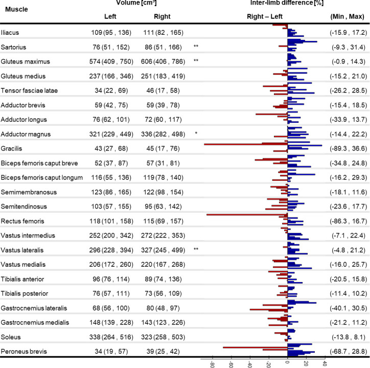
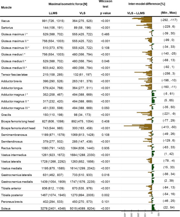
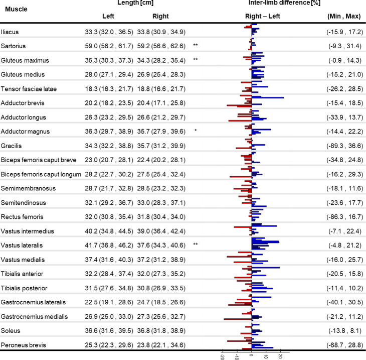
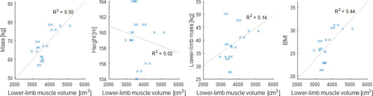
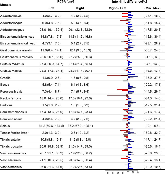
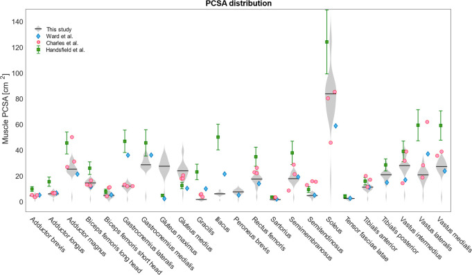
The ability of muscles to produce force depends, among others, on their anatomical features and it is altered by ageing-associated weakening. However, a clear characterisation of these features, highly relevant for older individuals, is still lacking. This study hence aimed at characterising muscle volume, length, and physiological cross-sectional area (PCSA) and their variability, between body sides and between individuals, in a group of post-menopausal women. Lower-limb magnetic resonance images were acquired from eleven participants (69 (7) y. o., 66.9 (7.7) kg, 159 (3) cm). Twenty-three muscles were manually segmented from the images and muscle volume, length and PCSA were calculated from this dataset. Personalised maximal isometric force was then calculated using the latter information. The percentage difference between the muscles of the two lower limbs was up to 89% and 22% for volume and length, respectively, and up to 84% for PCSA, with no recognisable pattern associated with limb dominance. Between-subject coefficients of variation reached 36% and 13% for muscle volume and length, respectively. Generally, muscle parameters were similar to previous literature, but volumes were smaller than those from in-vivo young adults and slightly higher than ex-vivo ones. Maximal isometric force was found to be on average smaller than those obtained from estimates based on linear scaling of ex-vivo-based literature values. In conclusion, this study quantified for the first time anatomical asymmetry of lower-limb muscles in older women, suggesting that symmetry should not be assumed in this population. Furthermore, we showed that a scaling approach, widely used in musculoskeletal modelling, leads to an overestimation of the maximal isometric force for most muscles. This heavily questions the validity of this approach for older populations. As a solution, the unique dataset of muscle segmentation made available with this paper could support the development of alternative population-based scaling approaches, together with that of automatic tools for muscle segmentation.
肌肉产生力量的能力取决于其解剖特征,随着年龄的增长,这些特征会逐渐减弱。然而,对于老年人来说,这些特征的明确特征仍然缺乏。因此,本研究旨在描述一组绝经后女性的下肢肌肉体积、长度和生理横截面积(PCSA)及其变异性,包括身体两侧和个体之间的差异。从 11 名参与者(69(7)岁,66.9(7.7)kg,159(3)cm)获得下肢磁共振图像。从图像中手动分割出 23 块肌肉,并从该数据集计算出肌肉体积、长度和 PCSA。然后使用这些信息计算个性化的最大等长力。两条下肢肌肉的差异百分比高达 89%和 22%,分别为体积和长度,PCSA 高达 84%,且无明显与肢体优势相关的模式。个体间变异系数分别达到 36%和 13%,用于肌肉体积和长度。一般来说,肌肉参数与之前的文献相似,但体积比活体年轻人小,略高于体外的。最大等长力平均小于基于体外文献值线性缩放估计的那些。总之,本研究首次定量描述了老年女性下肢肌肉的解剖学不对称性,表明在该人群中不应假设对称性。此外,我们表明,广泛用于肌肉骨骼建模的缩放方法会导致大多数肌肉的最大等长力高估。这严重质疑了该方法在老年人群中的有效性。作为一种解决方案,本文提供的肌肉分割独特数据集可以支持替代基于人群的缩放方法的开发,以及用于肌肉分割的自动工具的开发。