• 文献检索
  • 文档翻译
  • 深度研究
  • 学术资讯
  • Suppr Zotero 插件Zotero 插件
  • 邀请有礼
  • 套餐&价格
  • 历史记录
应用&插件
Suppr Zotero 插件Zotero 插件浏览器插件Mac 客户端Windows 客户端微信小程序
定价
高级版会员购买积分包购买API积分包
服务
文献检索文档翻译深度研究API 文档MCP 服务
关于我们
关于 Suppr公司介绍联系我们用户协议隐私条款
关注我们

Suppr 超能文献

核心技术专利:CN118964589B侵权必究
粤ICP备2023148730 号-1Suppr @ 2026

文献检索

告别复杂PubMed语法,用中文像聊天一样搜索,搜遍4000万医学文献。AI智能推荐,让科研检索更轻松。

立即免费搜索

文件翻译

保留排版,准确专业,支持PDF/Word/PPT等文件格式,支持 12+语言互译。

免费翻译文档

深度研究

AI帮你快速写综述,25分钟生成高质量综述,智能提取关键信息,辅助科研写作。

立即免费体验

多平台成像在癌症动物模型中的临床前应用

Preclinical Applications of Multi-Platform Imaging in Animal Models of Cancer.

作者信息

Serkova Natalie J, Glunde Kristine, Haney Chad R, Farhoud Mohammed, De Lille Alexandra, Redente Elizabeth F, Simberg Dmitri, Westerly David C, Griffin Lynn, Mason Ralph P

机构信息

Department of Radiology, University of Colorado Anschutz Medical Campus, Aurora, Colorado.

Animal Imaging Shared Resource, University of Colorado Cancer Center, Aurora, Colorado.

出版信息

Cancer Res. 2021 Mar 1;81(5):1189-1200. doi: 10.1158/0008-5472.CAN-20-0373. Epub 2020 Dec 1.

DOI:10.1158/0008-5472.CAN-20-0373
PMID:33262127
原文链接:https://pmc.ncbi.nlm.nih.gov/articles/PMC8026542/
Abstract

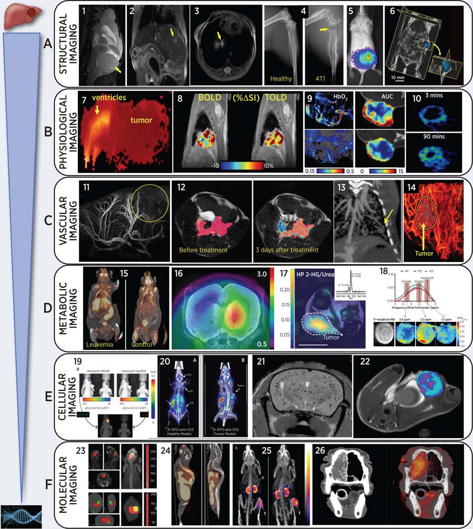
In animal models of cancer, oncologic imaging has evolved from a simple assessment of tumor location and size to sophisticated multimodality exploration of molecular, physiologic, genetic, immunologic, and biochemical events at microscopic to macroscopic levels, performed noninvasively and sometimes in real time. Here, we briefly review animal imaging technology and molecular imaging probes together with selected applications from recent literature. Fast and sensitive optical imaging is primarily used to track luciferase-expressing tumor cells, image molecular targets with fluorescence probes, and to report on metabolic and physiologic phenotypes using smart switchable luminescent probes. MicroPET/single-photon emission CT have proven to be two of the most translational modalities for molecular and metabolic imaging of cancers: immuno-PET is a promising and rapidly evolving area of imaging research. Sophisticated MRI techniques provide high-resolution images of small metastases, tumor inflammation, perfusion, oxygenation, and acidity. Disseminated tumors to the bone and lung are easily detected by microCT, while ultrasound provides real-time visualization of tumor vasculature and perfusion. Recently available photoacoustic imaging provides real-time evaluation of vascular patency, oxygenation, and nanoparticle distributions. New hybrid instruments, such as PET-MRI, promise more convenient combination of the capabilities of each modality, enabling enhanced research efficacy and throughput.

摘要

在癌症动物模型中,肿瘤成像已从对肿瘤位置和大小的简单评估发展到对微观到宏观层面的分子、生理、遗传、免疫和生化事件进行复杂的多模态探索,且以非侵入性方式进行,有时甚至是实时的。在此,我们简要回顾动物成像技术和分子成像探针,并结合近期文献中的选定应用进行阐述。快速且灵敏的光学成像主要用于追踪表达荧光素酶的肿瘤细胞、用荧光探针成像分子靶点,以及使用智能可切换发光探针报告代谢和生理表型。微型正电子发射断层扫描/单光子发射计算机断层扫描已被证明是癌症分子和代谢成像中最具转化性的两种模态:免疫正电子发射断层扫描是一个有前景且发展迅速的成像研究领域。先进的磁共振成像技术可提供小转移灶、肿瘤炎症、灌注、氧合和酸度的高分辨率图像。微型计算机断层扫描能轻松检测出扩散至骨骼和肺部的肿瘤,而超声可实时显示肿瘤血管系统和灌注情况。最近出现的光声成像可对血管通畅性、氧合和纳米颗粒分布进行实时评估。新型混合仪器,如正电子发射断层扫描 - 磁共振成像,有望更便捷地结合每种模态的功能,提高研究效率和通量。

https://cdn.ncbi.nlm.nih.gov/pmc/blobs/28c0/8026542/df754496f6bc/nihms-1651747-f0001.jpg

相似文献

1
Preclinical Applications of Multi-Platform Imaging in Animal Models of Cancer.多平台成像在癌症动物模型中的临床前应用
Cancer Res. 2021 Mar 1;81(5):1189-1200. doi: 10.1158/0008-5472.CAN-20-0373. Epub 2020 Dec 1.
2
PET-MR and SPECT-MR multimodality probes: Development and challenges.正电子发射断层扫描磁共振成像(PET-MR)和单光子发射计算机断层扫描磁共振成像(SPECT-MR)多模态探针:开发与挑战。
Theranostics. 2018 Nov 29;8(22):6210-6232. doi: 10.7150/thno.26610. eCollection 2018.
3
Discrepancies and priorities in staging and restaging malignant lymphoma by SPET, SPET/CT, PET/CT and PET/MRI.利用单光子发射计算机断层扫描(SPET)、SPET/CT、PET/CT和PET/MRI对恶性淋巴瘤进行分期及再分期时的差异与优先事项。
Hell J Nucl Med. 2013 Sep-Dec;16(3):223-9. doi: 10.1967/s002449910098. Epub 2013 Oct 2.
4
Positron emission tomography-magnetic resonance imaging as a research tool in musculoskeletal conditions.正电子发射断层扫描-磁共振成像作为肌肉骨骼疾病的研究工具。
Q J Nucl Med Mol Imaging. 2022 Mar;66(1):15-30. doi: 10.23736/S1824-4785.22.03434-3. Epub 2022 Jan 10.
5
Review of functional/anatomical imaging in oncology.肿瘤学中功能/解剖成像综述。
Nucl Med Commun. 2012 Apr;33(4):349-61. doi: 10.1097/MNM.0b013e32834ec8a5.
6
Current and future trends in multimodality imaging of coronary artery disease.冠状动脉疾病多模态成像的当前及未来趋势
Expert Rev Cardiovasc Ther. 2015 Jun;13(6):715-31. doi: 10.1586/14779072.2015.1039991. Epub 2015 Apr 25.
7
Oncologic imaging end-points for the assessment of therapy response.用于评估治疗反应的肿瘤影像学终点。
Recent Pat Anticancer Drug Discov. 2009 Jan;4(1):36-53. doi: 10.2174/157489209787002434.
8
More advantages in detecting bone and soft tissue metastases from prostate cancer using F-PSMA PET/CT.使用F-PSMA PET/CT检测前列腺癌骨和软组织转移方面有更多优势。
Hell J Nucl Med. 2019 Jan-Apr;22(1):6-9. doi: 10.1967/s002449910952. Epub 2019 Mar 7.
9
Whole-body magnetic resonance imaging and positron emission tomography-computed tomography in oncology.肿瘤学中的全身磁共振成像和正电子发射断层扫描-计算机断层扫描
Top Magn Reson Imaging. 2007 Jun;18(3):193-202. doi: 10.1097/RMR.0b013e318093e6bo.
10
The Novel DPP-BDT Nanoparticles as Efficient Photoacoustic Imaging and Positron Emission Tomography Agents in Living Mice.新型 DPP-BDT 纳米颗粒作为活体小鼠高效的光声成像和正电子发射断层成像剂。
Int J Nanomedicine. 2020 Jul 14;15:5017-5026. doi: 10.2147/IJN.S238679. eCollection 2020.

引用本文的文献

1
A pair of congenic mice for imaging of transplants by positron emission tomography using anti-transferrin receptor nanobodies.一对用于通过使用抗转铁蛋白受体纳米抗体的正电子发射断层扫描对移植进行成像的同类系小鼠。
Elife. 2025 Aug 18;14:RP104302. doi: 10.7554/eLife.104302.
2
Large animal models in gynecology: status and future perspectives.妇科领域的大型动物模型:现状与未来展望
Front Vet Sci. 2025 May 7;12:1588098. doi: 10.3389/fvets.2025.1588098. eCollection 2025.
3
Evaluating Therapeutic Efficacy of the Vascular Disrupting Agent OXi8007 Against Kidney Cancer in Mice.

本文引用的文献

1
Evaluating online filtering algorithms to enhance dynamic multispectral optoacoustic tomography.评估在线滤波算法以增强动态多光谱光声断层成像。
Photoacoustics. 2020 May 16;19:100184. doi: 10.1016/j.pacs.2020.100184. eCollection 2020 Sep.
2
Bioreductively Activatable Prodrug Conjugates of Combretastatin A-1 and Combretastatin A-4 as Anticancer Agents Targeted toward Tumor-Associated Hypoxia.作为靶向肿瘤相关缺氧的抗癌剂的康普瑞汀A-1和康普瑞汀A-4的生物还原可激活前药缀合物。
J Nat Prod. 2020 Apr 24;83(4):937-954. doi: 10.1021/acs.jnatprod.9b00773. Epub 2020 Mar 20.
3
Preclinical Imaging Using Single Track Location Shear Wave Elastography: Monitoring the Progression of Murine Pancreatic Tumor Liver Metastasis In Vivo.
评估血管破坏剂OXi8007对小鼠肾癌的治疗效果。
Cancers (Basel). 2025 Feb 24;17(5):771. doi: 10.3390/cancers17050771.
4
Advancements in molecular imaging probes for precision diagnosis and treatment of prostate cancer.用于前列腺癌精准诊断与治疗的分子成像探针的进展。
J Zhejiang Univ Sci B. 2025 Jan 15;26(2):124-144. doi: 10.1631/jzus.B2300614.
5
Molecular effects of clinically relevant chemotherapeutic agents on choline phospholipid metabolism in triple negative breast cancer cells.临床相关化疗药物对三阴性乳腺癌细胞胆碱磷脂代谢的分子效应
Transl Oncol. 2025 Mar;53:102311. doi: 10.1016/j.tranon.2025.102311. Epub 2025 Feb 7.
6
A Vector Nanoplatform for the Bioimaging of Deep-Seated Tumors.用于深部肿瘤生物成像的载体纳米平台
Acta Naturae. 2024 Apr-Jun;16(2):72-81. doi: 10.32607/actanaturae.27425.
7
Bacterial imaging in tumour diagnosis.肿瘤诊断中的细菌成像。
Microb Biotechnol. 2024 Jun;17(6):e14474. doi: 10.1111/1751-7915.14474.
8
High-Spatial-Resolution Benchtop X-ray Fluorescence Imaging through Bragg-Diffraction-Based Focusing with Bent Mosaic Graphite Crystals: A Simulation Study.基于弯曲镶嵌石墨晶体的布拉格衍射聚焦的高空间分辨率台式 X 射线荧光成像:模拟研究。
Int J Mol Sci. 2024 Apr 26;25(9):4733. doi: 10.3390/ijms25094733.
9
Orthotopic and metastatic tumour models in preclinical cancer research.临床前癌症研究中的原位和转移瘤模型。
Pharmacol Ther. 2024 May;257:108631. doi: 10.1016/j.pharmthera.2024.108631. Epub 2024 Mar 11.
10
Preclinical magnetic resonance imaging and spectroscopy in the fields of radiological technology, medical physics, and radiology.放射技术、医学物理和放射学领域的临床前磁共振成像和光谱学。
Radiol Phys Technol. 2024 Mar;17(1):47-59. doi: 10.1007/s12194-024-00785-y. Epub 2024 Feb 14.
基于单轨迹定位剪切波弹性成像的临床前影像学研究:活体监测小鼠胰腺肿瘤肝转移的进展。
IEEE Trans Med Imaging. 2020 Jul;39(7):2426-2439. doi: 10.1109/TMI.2020.2971422. Epub 2020 Feb 3.
4
Ultra-early changes in vascular parameters from dynamic contrast enhanced MRI of breast cancer xenografts following systemic therapy with doxorubicin and liver X receptor agonist.系统给予阿霉素和肝 X 受体激动剂治疗后,乳腺癌移植瘤的动态对比增强 MRI 中血管参数的超早期变化。
Cancer Imaging. 2019 Dec 19;19(1):88. doi: 10.1186/s40644-019-0280-y.
5
BPTF regulates growth of adult and pediatric high-grade glioma through the MYC pathway.BPTF 通过 MYC 通路调控成人和儿童高级别脑胶质瘤的生长。
Oncogene. 2020 Mar;39(11):2305-2327. doi: 10.1038/s41388-019-1125-7. Epub 2019 Dec 16.
6
Metabolic Imaging Using Hyperpolarized Pyruvate-Lactate Exchange Assesses Response or Resistance to the EGFR Inhibitor Cetuximab in Patient-Derived HNSCC Xenografts.利用超极化丙酮酸-乳酸交换进行代谢成像评估表皮生长因子受体抑制剂西妥昔单抗在患者来源的头颈部鳞状细胞癌异种移植物中的反应或耐药性。
Clin Cancer Res. 2020 Apr 15;26(8):1932-1943. doi: 10.1158/1078-0432.CCR-19-1369. Epub 2019 Dec 12.
7
ImmunoPET, [Cu]Cu-DOTA-Anti-CD33 PET-CT, Imaging of an AML Xenograft Model.免疫 PET(ImmunoPET),[Cu]Cu-DOTA-Anti-CD33 PET-CT,AML 异种移植模型的成像。
Clin Cancer Res. 2019 Dec 15;25(24):7463-7474. doi: 10.1158/1078-0432.CCR-19-1106. Epub 2019 Sep 23.
8
Differentiating lung cancer and infection based on measurements of extracellular pH with acidoCEST MRI.利用 acidoCEST MRI 测量细胞外 pH 值区分肺癌和感染。
Sci Rep. 2019 Sep 10;9(1):13002. doi: 10.1038/s41598-019-49514-1.
9
On the promise of artificial intelligence for standardizing radiographic response assessment in gliomas.关于人工智能在标准化神经胶质瘤放射学反应评估方面的前景。
Neuro Oncol. 2019 Nov 4;21(11):1346-1347. doi: 10.1093/neuonc/noz162.
10
Quantitative Drug Release Monitoring in Tumors of Living Subjects by Magnetic Particle Imaging Nanocomposite.通过磁粒子成像纳米复合材料在活体研究对象肿瘤中进行定量药物释放监测。
Nano Lett. 2019 Oct 9;19(10):6725-6733. doi: 10.1021/acs.nanolett.9b01202. Epub 2019 Sep 25.