Columbia University PET Center, Department of Radiology, Columbia University Medical Center, New York, NY, USA.
Department of Biostatistics and Data Science, Division of Public Health Sciences, Wake Forest School of Medicine, Winston-Salem, NC, USA.
Drug Deliv. 2020 Dec;27(1):1686-1694. doi: 10.1080/10717544.2020.1833381.
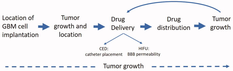
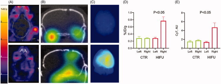
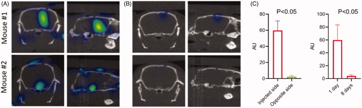
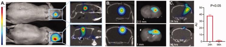
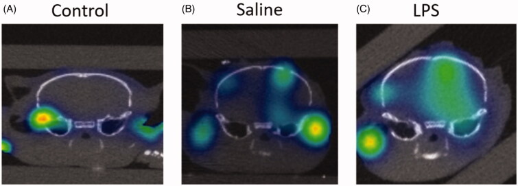
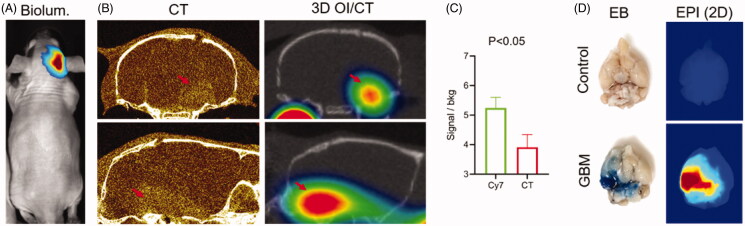
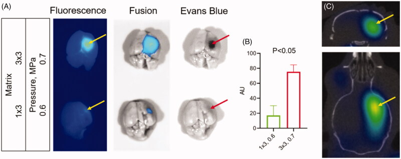
Multimodality 3D Optical Imaging (OI)/CT has the potential to play a major role in drug development for glioblastomas (GBM), as it is an accessible preclinical method. To demonstrate the potential of 3D OI/CT to visualize orthotopic GBM implantation, we labeled GBM cells with Cy7 and imaged their location using 3D OI/CT. To confirm the accuracy of the spatial localization and demonstrate the ability to image locoregionally delivered therapies, we labeled mouse albumin with Cy7 (Cy7ALB) and delivered it via locoregional infusion 1 mm or 3 mm into the brain and demonstrated correlation of signal between the 3D OI/CT and post necropsy brain slices. In addition, we demonstrated the potential of systemically delivered Cy7ALB contrast to detect blood-brain barrier (BBB) permeability caused by orthotopic GBMs using 3D OI/CT. We also tested the potential of 3D OI/CT to assess focal BBB permeability induced by high intensity focused ultrasound (HIFU), a methodology being used in clinical trials to noninvasively permeabilize the BBB for systemic therapeutic delivery to GBM. We demonstrated the ability of systemic Cy7ALB contrast together with 3D OI/CT to accurately assess real-time HIFU-induced BBB permeability, which correlated to post necropsy imaging of brains. Furthermore, we demonstrated that 3D OI/CT can also image the therapeutic distribution of a Cy7-labeled anti-PD-1 antibody, a prototype translational antibody therapy. We successfully imaged real-time antibody distribution after HIFU-induced BBB permeability, which correlated with post necropsy Cy7 signal and translational PET imaging after injection of [Zr] anti-PD-1 antibody. Thus, we demonstrated the broad potential of using 3D OI/CT as an accessible preclinical tool to develop anti-GBM therapies.
多模态 3D 光学成像 (OI)/CT 有可能在胶质母细胞瘤 (GBM) 的药物开发中发挥重要作用,因为它是一种易于获得的临床前方法。为了证明 3D OI/CT 可视化原位 GBM 植入的潜力,我们用 Cy7 标记 GBM 细胞,并使用 3D OI/CT 对其位置进行成像。为了确认空间定位的准确性并证明对局部递送疗法进行成像的能力,我们用 Cy7 标记小鼠白蛋白(Cy7ALB),并通过局部输注将其递送至脑内 1 毫米或 3 毫米处,证明了 3D OI/CT 和死后脑切片之间的信号相关性。此外,我们还展示了全身性递送 Cy7ALB 对比剂通过 3D OI/CT 检测由原位 GBM 引起的血脑屏障 (BBB) 通透性的潜力。我们还测试了 3D OI/CT 评估高强度聚焦超声 (HIFU) 诱导的局灶性 BBB 通透性的潜力,该方法正在临床试验中用于非侵入性地使 BBB 通透性增加,以便将系统治疗药物递送至 GBM。我们证明了全身性 Cy7ALB 对比剂与 3D OI/CT 一起准确评估实时 HIFU 诱导的 BBB 通透性的能力,这与死后脑成像相关。此外,我们还证明 3D OI/CT 还可以对 Cy7 标记的抗 PD-1 抗体(一种翻译抗体治疗的原型)的治疗分布进行成像。我们成功地对 HIFU 诱导的 BBB 通透性后的实时抗体分布进行了成像,这与死后的 Cy7 信号以及注射 [Zr] 抗 PD-1 抗体后的翻译 PET 成像相关。因此,我们证明了使用 3D OI/CT 作为易于获得的临床前工具来开发抗 GBM 疗法的广泛潜力。