Cedars-Sinai Medical Center, Department of Biomedical Sciences, Biomedical Imaging Research Institute, Los Angeles, CA, United States of America.
Department of Bioengineering, University of California, Los Angeles, CA, United States of America.
PLoS One. 2020 Dec 2;15(12):e0243207. doi: 10.1371/journal.pone.0243207. eCollection 2020.
Intramyocardial hemorrhage following reperfusion is strongly associated with major adverse cardiovascular events in myocardial infarction (MI) patients; yet the mechanisms contributing to these outcomes are not well understood. Large animal models have been used to investigate intramyocardial hemorrhage, but they are exorbitantly expensive and difficult to use for mechanistic studies. In contrast, rat models are widely used to investigate mechanistic aspects of cardiovascular physiology, but a rat model that consistently recapitulates the characteristics of an hemorrhagic MI does not exist. To bridge this gap, we investigated the physiological conditions of MI that would create intramyocardial hemorrhage in rats so that a reliable model of hemorrhagic MI would become available for basic research.
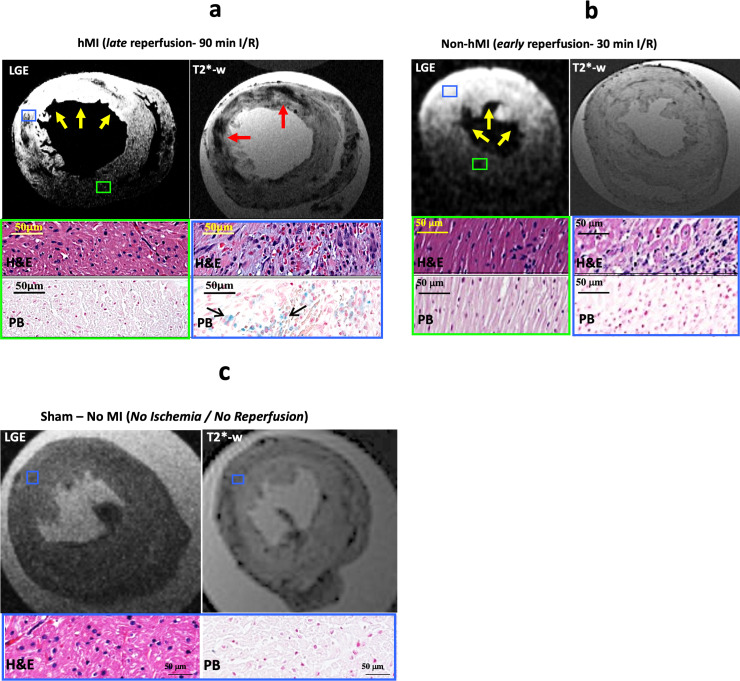
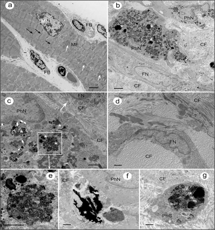
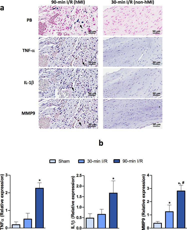
METHODS & RESULTS: Sprague-Dawley rats underwent either a 90-minute (90-min) ischemia and then reperfusion (I/R) (n = 22) or 30-minute (30-min) I/R (n = 18) of the left anterior descending coronary artery. Sham rats (n = 12) were used as controls. 90-min I/R consistently yielded hemorrhagic MI, while 30-min I/R consistently yielded non-hemorrhagic MI. Twenty-four hours post-reperfusion, ex-vivo late-gadolinium-enhancement (LGE) and T2* cardiac MRI performed on excised hearts from 90-min I/R rats revealed colocalization of iron deposits within the scarred tissue; however, in 30-min I/R rats scar was evident on LGE but no evidence of iron was found on T2* CMR. Histological studies verified tissue damage (H&E) detected on LGE and the presence of iron (Perl's stain) observed on T2*-CMR. At week 4 post-reperfusion, gene and protein expression of proinflammatory markers (TNF-α, IL-1β and MMP-9) were increased in the 90-min I/R group when compared to 30-min I/R groups. Further, transmission electron microscopy performed on 90-min I/R myocardium that were positive for iron on T2* CMR and Perl's stain showed accumulation of granular iron particles within the phagosomes.
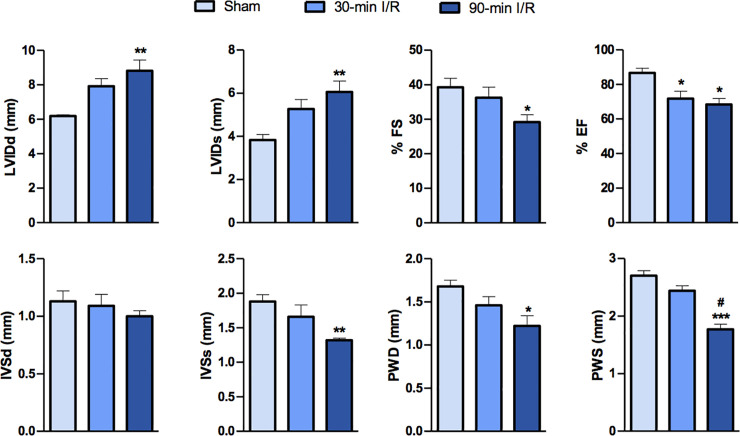
Ischemic time prior to reperfusion is a critical factor in determining whether a MI is hemorrhagic or non-hemorrhagic in rats. Specifically, a period of 90-min of ischemia prior to reperfusion can produce rat models of hemorrhagic MI, while 30-minutes of ischemia prior to reperfusion can ensure that the MIs are non-hemorrhagic. Hemorrhagic MIs in rats result in marked increase in iron deposition, proinflammatory burden and adverse left-ventricular remodeling compared to rats with non-hemorrhagic MIs.
心肌再灌注后发生的心肌内出血与心肌梗死(MI)患者的主要不良心血管事件密切相关;然而,导致这些结果的机制尚不清楚。大型动物模型已被用于研究心肌内出血,但它们非常昂贵,并且难以用于机制研究。相比之下,大鼠模型被广泛用于研究心血管生理学的机制方面,但不存在一种能够一致重现出血性 MI 特征的大鼠模型。为了弥补这一差距,我们研究了 MI 的生理条件,这些条件会导致大鼠的心肌内出血,以便为基础研究提供一种可靠的出血性 MI 模型。
Sprague-Dawley 大鼠接受左前降支冠状动脉 90 分钟(90 分钟)缺血再灌注(I/R)(n = 22)或 30 分钟(30 分钟)I/R(n = 18)。假手术大鼠(n = 12)用作对照。90 分钟 I/R 始终导致出血性 MI,而 30 分钟 I/R 始终导致非出血性 MI。再灌注后 24 小时,对 90 分钟 I/R 大鼠切除心脏进行的离体晚期钆增强(LGE)和 T2心脏 MRI 显示,铁沉积物在疤痕组织内的定位一致;然而,在 30 分钟 I/R 大鼠中,LGE 显示出疤痕,但在 T2CMR 上未发现铁的存在。组织学研究证实了 LGE 检测到的组织损伤(H&E)和 T2*-CMR 上观察到的铁(Perl 染色)的存在。再灌注后 4 周,与 30 分钟 I/R 组相比,90 分钟 I/R 组促炎标志物(TNF-α、IL-1β 和 MMP-9)的基因和蛋白表达增加。此外,对 T2*CMR 和 Perl 染色呈阳性的 90 分钟 I/R 心肌进行透射电子显微镜检查显示,吞噬体中积累了颗粒状铁颗粒。
缺血再灌注前的缺血时间是决定大鼠 MI 是否为出血性或非出血性的关键因素。具体来说,缺血 90 分钟再灌注可导致大鼠出血性 MI 模型,而缺血 30 分钟再灌注可确保 MI 为非出血性。与非出血性 MI 大鼠相比,大鼠出血性 MI 导致铁沉积、促炎负担和左心室重构的明显增加。