Polaris, Imaging group, Dept IICD, University of Sheffield, Sheffield, UK.
Academic Directorate of Respiratory Medicine, Sheffield Teaching Hospitals NHS Foundation Trust, Sheffield, Sheffield, UK.
Thorax. 2021 Feb;76(2):144-151. doi: 10.1136/thoraxjnl-2019-214375. Epub 2020 Dec 3.
Idiopathic pulmonary fibrosis (IPF) is a fatal disease of lung scarring. Many patients later develop raised pulmonary vascular pressures, sometimes disproportionate to the interstitial disease. Previous therapeutic approaches that have targeted pulmonary vascular changes have not demonstrated clinical efficacy, and quantitative assessment of regional pulmonary vascular involvement using perfusion imaging may provide a biomarker for further therapeutic insights.
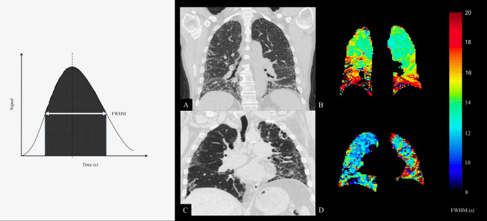
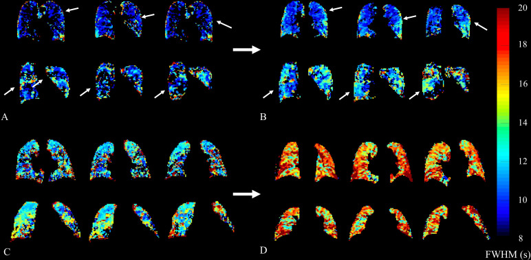
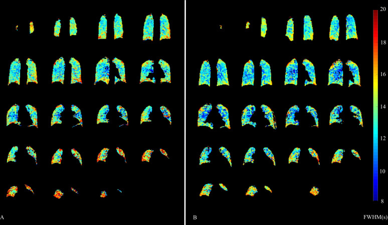
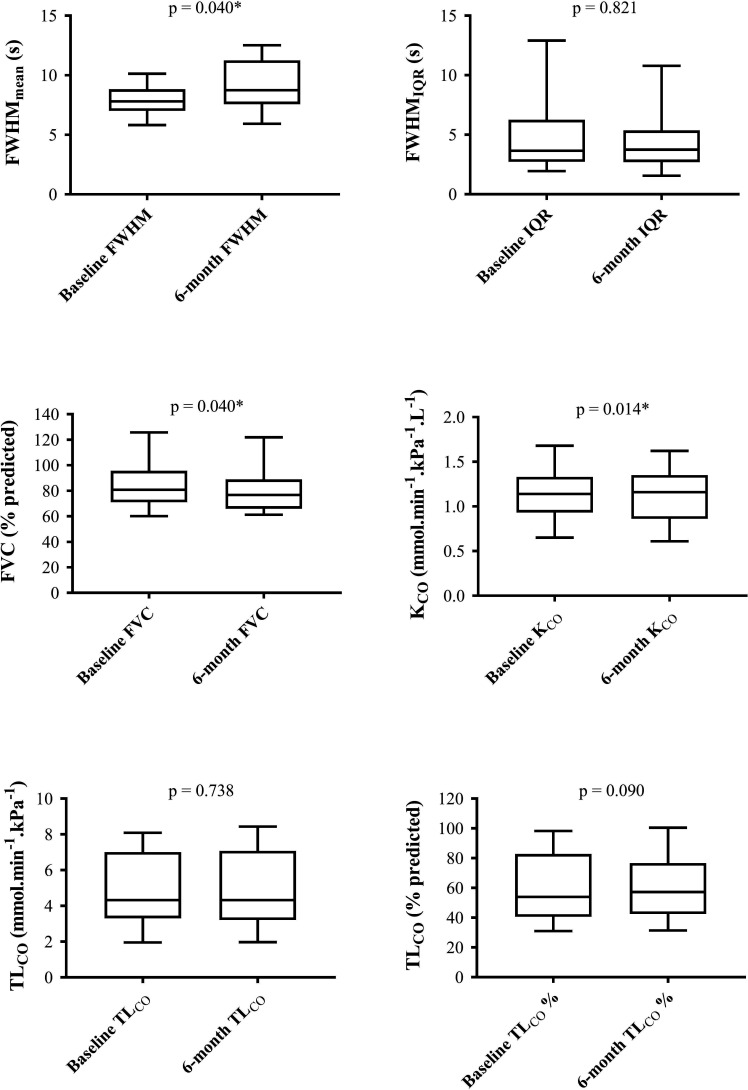
We studied 23 participants with IPF, using dynamic contrast-enhanced MRI (DCE-MRI) and pulmonary function tests, including forced vital capacity (FVC), transfer factor (TL) and coefficient (K) of the lungs for carbon monoxide. DCE-MRI parametric maps were generated including the full width at half maximum (FWHM) of the bolus transit time through the lungs. Key metrics used were mean (FWHM) and heterogeneity (FWHM). Nineteen participants returned at 6 months for repeat assessment.
Spearman correlation coefficients were identified between TL and FWHM (r=-0.46; p=0.026), K and FWHM (r=-0.42; p=0.047) and K and FWHM (r=-0.51; p=0.013) at baseline. No statistically significant correlations were seen between FVC and DCE-MRI metrics. Follow-up at 6 months demonstrated statistically significant decline in FVC (p=0.040) and K (p=0.014), with an increase in FWHM (p=0.040), but no significant changes in TL (p=0.090) nor FWHM (p=0.821).
DCE-MRI first pass perfusion demonstrates correlations with existing physiological gas exchange metrics, suggesting that capillary perfusion deficit (as well as impaired interstitial diffusion) may contribute to gas exchange limitation in IPF. FWHM showed a significant increase over a 6-month period and has potential as a quantitative biomarker of pulmonary vascular disease progression in IPF.
特发性肺纤维化(IPF)是一种致命的肺部瘢痕疾病。许多患者后来会出现肺动脉压升高,有时与间质疾病不成比例。以前针对肺血管变化的治疗方法并未显示出临床疗效,使用灌注成像对区域性肺血管受累进行定量评估可能为进一步的治疗提供生物标志物。
我们研究了 23 名 IPF 患者,使用动态对比增强 MRI(DCE-MRI)和肺功能测试,包括用力肺活量(FVC)、转移因子(TL)和一氧化碳肺系数(K)。生成了包括通过肺部的 bolus 传输时间的半最大值全宽(FWHM)在内的 DCE-MRI 参数图。使用的关键指标是平均值(FWHM)和异质性(FWHM)。19 名参与者在 6 个月时返回进行重复评估。
在基线时,TL 与 FWHM 之间存在 Spearman 相关系数(r=-0.46;p=0.026),K 与 FWHM 之间存在相关系数(r=-0.42;p=0.047),K 与 FWHM 之间存在相关系数(r=-0.51;p=0.013)。FVC 与 DCE-MRI 指标之间未见统计学显著相关性。6 个月的随访显示 FVC(p=0.040)和 K(p=0.014)有统计学显著下降,FWHM 增加(p=0.040),但 TL(p=0.090)和 FWHM(p=0.821)无显著变化。
DCE-MRI 首次通过灌注显示与现有的生理气体交换指标相关,表明毛细血管灌注不足(以及间质扩散受损)可能导致 IPF 中的气体交换受限。FWHM 在 6 个月内显著增加,具有作为 IPF 中肺血管疾病进展的定量生物标志物的潜力。