Singh Srishti, Raghavan Bagyam, Geethapriya Sivaramalingam, Sathyasree V V, Govindaraj Jayaraj, Padmanabhan G, Krishna Murali, Arafath Rasheed
Department of Radiodiagnosis, Apollo Speciality Hospital, Chennai, Tamil Nadu, India.
Indian J Radiol Imaging. 2020 Jul-Sep;30(3):319-326. doi: 10.4103/ijri.IJRI_59_20. Epub 2020 Oct 15.
Prognosis and survival rates for breast cancer vary greatly depending on the cancer stage of the patient. Instead of a step-by-step approach using multiple investigations, we can get all the information about the metastatic load of the disease in PET-CT imaging by one single investigation. There is also a correlation between prognosis, FDG uptake, and molecular subtype of breast cancer (Luminal A, Luminal B, Human epidermal growth factor receptor 2 (HER2) positive and Triple-negative). Pre-treatment baseline PET-CT scan was done in 156 unilateral early and operable breast cancer patients from November 2017 to April 2019 in our prospective observational study.
To evaluate the utility of PET-CT in staging and upstaging of early and operable breast cancer by detection of unsuspected lymph nodes and distant organ metastases.To determine the prognostic association between SUVmax of the primary breast lesion in the upstaged cases and the molecular subtypes.
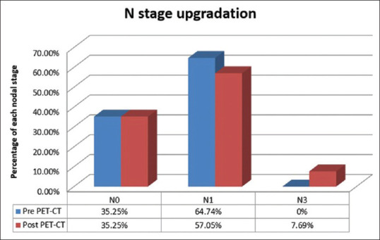
Thus, PET-CT can serve as one-stop imaging in unilateral operable early breast cancer patients for upstaging and prognostication based on the correlation of SUVmax with molecular subtypes of breast lesions in patients who will surely benefit from whole-body imaging.Out of 156 patients, approximately 27 patients were upstaged after pre-treatment PET CT.Six patients were upstaged to stage IIIC and 21 patients were upstaged to IV.Regional nodes like internal mammary and supraclavicular nodes were detected in 7 patients and 5 patients, respectively, out of 156 patients.Non-regional distant nodes and organ metastases were detected in 11 and 18 patients out of 156 patients.Most common molecular subtype detected in the upstaged cases in our study was Luminal A (13 patients) followed by Triple negative (6), Luminal B (3) and HER2-neu-positive subtypes (1).
FDG PET-CT is a substantial modality to provide information on regional, non regional lymph nodes and distant metastases in early operable breast cancer.It helps in evaluating the whole body metastatic burden in a single sitting, therefore, reducing the need for multiple investigations.SUVmax association of the index lesion with molecular subtype in the FDG PET scanning can serve as a prognostication factor in operable early breast cancer patients.
乳腺癌的预后和生存率因患者的癌症分期不同而有很大差异。我们无需采用多项检查的分步方法,通过一次PET-CT检查就能获取有关疾病转移负荷的所有信息。乳腺癌的预后、氟代脱氧葡萄糖(FDG)摄取与分子亚型(管腔A型、管腔B型、人表皮生长因子受体2(HER2)阳性和三阴性)之间也存在关联。在我们的前瞻性观察研究中,于2017年11月至2019年4月对156例单侧早期可手术乳腺癌患者进行了治疗前基线PET-CT扫描。
通过检测未被怀疑的淋巴结和远处器官转移,评估PET-CT在早期可手术乳腺癌分期及再分期中的作用。确定再分期病例中原发性乳腺病变的最大标准摄取值(SUVmax)与分子亚型之间的预后关联。
因此,基于SUVmax与乳腺病变分子亚型的相关性,PET-CT可作为单侧可手术早期乳腺癌患者进行再分期和预后评估的一站式成像检查,这类患者肯定能从全身成像检查中获益。在156例患者中,约27例患者在治疗前PET-CT检查后进行了再分期。6例患者被重新分期为IIIC期,21例患者被重新分期为IV期。在156例患者中,分别有7例和5例检测到内乳和锁骨上淋巴结等区域淋巴结。在156例患者中,分别有11例和18例检测到非区域远处淋巴结和器官转移。在我们研究中,再分期病例中检测到的最常见分子亚型是管腔A型(13例),其次是三阴性(6例)、管腔B型(3例)和HER2-神经阳性亚型(1例)。
FDG PET-CT是一种重要的检查方式,可提供早期可手术乳腺癌区域、非区域淋巴结及远处转移的信息。它有助于在一次检查中评估全身转移负担,从而减少多项检查的需求。FDG PET扫描中指标病变的SUVmax与分子亚型的关联可作为可手术早期乳腺癌患者的预后因素。