• 文献检索
  • 文档翻译
  • 深度研究
  • 学术资讯
  • Suppr Zotero 插件Zotero 插件
  • 邀请有礼
  • 套餐&价格
  • 历史记录
应用&插件
Suppr Zotero 插件Zotero 插件浏览器插件Mac 客户端Windows 客户端微信小程序
定价
高级版会员购买积分包购买API积分包
服务
文献检索文档翻译深度研究API 文档MCP 服务
关于我们
关于 Suppr公司介绍联系我们用户协议隐私条款
关注我们

Suppr 超能文献

核心技术专利:CN118964589B侵权必究
粤ICP备2023148730 号-1Suppr @ 2026

文献检索

告别复杂PubMed语法,用中文像聊天一样搜索,搜遍4000万医学文献。AI智能推荐,让科研检索更轻松。

立即免费搜索

文件翻译

保留排版,准确专业,支持PDF/Word/PPT等文件格式,支持 12+语言互译。

免费翻译文档

深度研究

AI帮你快速写综述,25分钟生成高质量综述,智能提取关键信息,辅助科研写作。

立即免费体验

F-FDG PET/CT对雌激素受体(ER)表达比例低的新诊断乳腺癌患者有益吗?

Is F-FDG PET/CT Beneficial for Newly Diagnosed Breast Cancer Patients With Low Proportion of ER Expression?

作者信息

Liu Jiachen, Sun Runlu, Yin Yuping, Li Jingyan, Liu Xuming, Liu Sheng, Zhang Zhanlei, Hu Jieting, Wan Xiaoting, Zhang Hong

机构信息

Department of Nuclear Medicine, Sun Yat-sen Memorial Hospital, Sun Yat-sen University, Guangzhou, China.

Department of Cardiology, Sun Yat-sen Memorial Hospital, Sun Yat-sen University, Guangzhou, China.

出版信息

Front Oncol. 2021 Nov 4;11:755899. doi: 10.3389/fonc.2021.755899. eCollection 2021.

DOI:10.3389/fonc.2021.755899
PMID:34804947
原文链接:https://pmc.ncbi.nlm.nih.gov/articles/PMC8599817/
Abstract

OBJECTIVE

It is unclear whether the receptor status of breast malignancy or the proportion of receptors expression is useful in the interpretation of F-FDG PET/CT. This study's purpose was to analyze whether F-FDG PET/CT was valuable for helping newly diagnosed breast cancer patients find suspected or unsuspected metastasis lesions based on the proportion of receptors expression.

MATERIALS AND METHODS

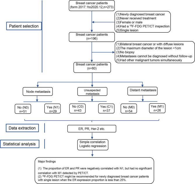
Eighty newly diagnosed breast cancer patients were divided into six groups, containing N0 (no extraaxillary lymph node metastasis), N1 (extraaxillary lymph node metastasis), M0 (no distant metastasis), and M1 (distant metastasis) groups, C0 (no unsuspected metastasis), and C1 (unsuspected metastasis and treatment plan changed) detected by PET/CT. The main data, including the proportion of receptors ER (estrogen receptor), PR (progesterone receptor), and Her-2 (human epidermal growth factor receptor 2) status, were extracted. Simple correlation and logistic regression were preformed to analyze the association between them.

RESULTS

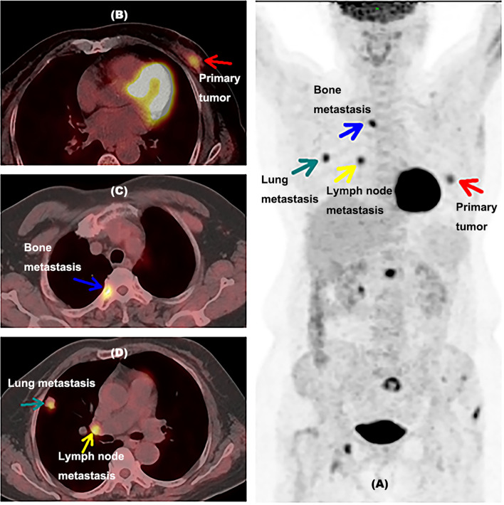
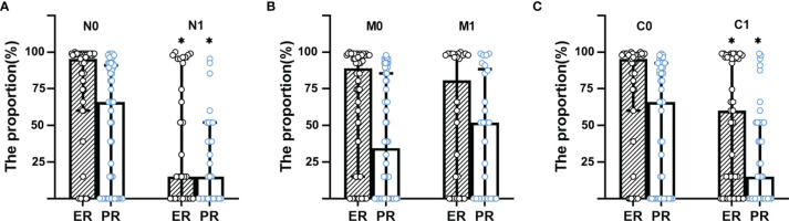
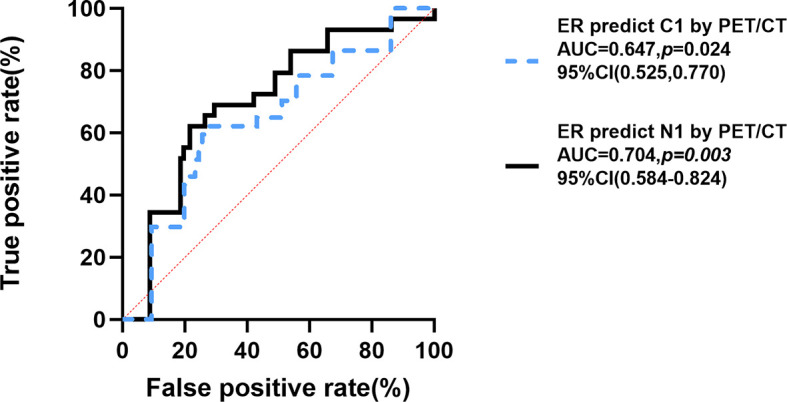
Patients in N1 group had lower proportion of ER (%) and PR (%) than that in N0 group (ER: 2 [0-80] 80 [15-95]; PR: 1 [0-10] 20 [0-45], <0.001). Moreover, the proportions of ER and PR were negatively correlated with N1 (ER: [r= -0.339, = 0.002], PR: [r= -0.247, = 0.011]) by simple correlation. Also, patients in C1 group had lower proportion of ER (%) and PR (%) than those in C0 group (ER: 10 [0-85] 80 [15-90], =0.026; PR: 1 [0-10] 20 [0-70], =0.041), while the distribution of ER and PR between M1 and M0 group had no significant difference. After the adjustment of traditional factors, the negative correlation between the proportion of ER (OR=0.986, 95% CI of OR [0.972-0.999], p=0.016) and C1 was found by logistic regression, cutoff value was 25% (ER) calculated by ROC (Receiver Operating Characteristic) curve (AUC [Area Under Curve]= 0.647, =0.024).

CONCLUSION

The proportion of ER in newly diagnosed breast cancer was negatively correlated with unsuspected metastasis detected by F-FDG PET/CT. F-FDG PET/CT might be recommended for newly diagnosed breast cancer patients with single lesions when the ER expression proportion is less than 25% to find unsuspected metastasis lesions and to modify treatment plan contrasted with conventional imaging and clinical examination.

摘要

目的

尚不清楚乳腺恶性肿瘤的受体状态或受体表达比例在F-FDG PET/CT的解读中是否有用。本研究的目的是基于受体表达比例分析F-FDG PET/CT对帮助新诊断的乳腺癌患者发现可疑或意外转移病灶是否有价值。

材料与方法

80例新诊断的乳腺癌患者被分为六组,包括N0(无腋窝外淋巴结转移)、N1(腋窝外淋巴结转移)、M0(无远处转移)和M1(远处转移)组,以及通过PET/CT检测出的C0(无意外转移)和C1(有意外转移且治疗方案改变)组。提取主要数据,包括雌激素受体(ER)、孕激素受体(PR)和人表皮生长因子受体2(Her-2)的受体状态比例。进行简单相关性分析和逻辑回归分析以分析它们之间的关联?

结果

N1组患者的ER(%)和PR(%)比例低于N0组(ER:2[0-80]对80[15-95];PR:1[0-10]对20[0-45],<0.001)。此外,通过简单相关性分析,ER和PR的比例与N1呈负相关(ER:[r = -0.339,P = 0.002],PR:[r = -0.247,P = 0.011])。同样,C1组患者的ER(%)和PR(%)比例低于C0组(ER:10[0-85]对80[15-90],P = 0.026;PR:1[0-10]对20[0-70],P = 0.041),而M1组和M0组之间ER和PR的分布无显著差异。在调整传统因素后,通过逻辑回归发现ER比例与C1之间存在负相关(OR = 0.986,OR的95%置信区间[0.972-0.999],P = 0.016),通过ROC(受试者工作特征)曲线计算的截断值为25%(ER)(曲线下面积[AUC]=0.647,P = 0.024)。

结论

新诊断乳腺癌中ER的比例与F-FDG PET/CT检测出的意外转移呈负相关。对于ER表达比例小于25%的新诊断的单发乳腺癌患者,可能推荐使用F-FDG PET/CT来发现意外转移病灶,并与传统影像学和临床检查相比修改治疗方案。

https://cdn.ncbi.nlm.nih.gov/pmc/blobs/3bce/8599817/8431e6af3226/fonc-11-755899-g005.jpg
https://cdn.ncbi.nlm.nih.gov/pmc/blobs/3bce/8599817/76993d06ae32/fonc-11-755899-g001.jpg
https://cdn.ncbi.nlm.nih.gov/pmc/blobs/3bce/8599817/e8f3ed517002/fonc-11-755899-g002.jpg
https://cdn.ncbi.nlm.nih.gov/pmc/blobs/3bce/8599817/2a9b81692255/fonc-11-755899-g003.jpg
https://cdn.ncbi.nlm.nih.gov/pmc/blobs/3bce/8599817/82a20268f624/fonc-11-755899-g004.jpg
https://cdn.ncbi.nlm.nih.gov/pmc/blobs/3bce/8599817/8431e6af3226/fonc-11-755899-g005.jpg

相似文献

1
Is F-FDG PET/CT Beneficial for Newly Diagnosed Breast Cancer Patients With Low Proportion of ER Expression?F-FDG PET/CT对雌激素受体(ER)表达比例低的新诊断乳腺癌患者有益吗?
Front Oncol. 2021 Nov 4;11:755899. doi: 10.3389/fonc.2021.755899. eCollection 2021.
2
F-FDG-PET/CT for systemic staging of patients with newly diagnosed ER-positive and HER2-positive breast cancer.氟代脱氧葡萄糖正电子发射断层扫描/计算机断层扫描在新发雌激素受体阳性和人表皮生长因子受体 2 阳性乳腺癌患者全身分期中的应用。
Eur J Nucl Med Mol Imaging. 2017 Aug;44(9):1420-1427. doi: 10.1007/s00259-017-3709-1. Epub 2017 Apr 29.
3
Correlation of breast cancer subtypes, based on estrogen receptor, progesterone receptor, and HER2, with functional imaging parameters from ⁶⁸Ga-RGD PET/CT and ¹⁸F-FDG PET/CT.基于雌激素受体、孕激素受体和人表皮生长因子受体2的乳腺癌亚型与⁶⁸Ga-RGD PET/CT及¹⁸F-FDG PET/CT功能成像参数的相关性
Eur J Nucl Med Mol Imaging. 2014 Aug;41(8):1534-43. doi: 10.1007/s00259-014-2744-4. Epub 2014 Mar 21.
4
F-FES PET/CT Influences the Staging and Management of Patients with Newly Diagnosed Estrogen Receptor-Positive Breast Cancer: A Retrospective Comparative Study with F-FDG PET/CT.F-FES PET/CT 对新诊断雌激素受体阳性乳腺癌患者分期和管理的影响:与 F-FDG PET/CT 的回顾性对比研究。
Oncologist. 2019 Dec;24(12):e1277-e1285. doi: 10.1634/theoncologist.2019-0096. Epub 2019 Jul 23.
5
The correlation of F-FDG PET/CT metabolic parameters, clinicopathological factors, and prognosis in breast cancer.乳腺癌中 F-FDG PET/CT 代谢参数与临床病理因素及预后的相关性。
Clin Transl Oncol. 2021 Mar;23(3):620-627. doi: 10.1007/s12094-020-02457-w. Epub 2020 Jul 18.
6
Diagnostic value of Ga-DOTATATE PET-CT imaging for staging of ER /PR HER2- breast cancer patients with metastatic disease: Comparison with conventional imaging with bone scan, diagnostic CT and F-FDG PET-CT in a prospective pilot trial.Ga-DOTATATE PET-CT 成像在 ER/PR HER2-转移性乳腺癌患者分期中的诊断价值:与骨扫描、诊断 CT 和 F-FDG PET-CT 常规成像的前瞻性先导试验比较。
J Med Imaging Radiat Oncol. 2022 Sep;66(6):731-737. doi: 10.1111/1754-9485.13342. Epub 2021 Oct 21.
7
(18)F-FDG-PET/CT for systemic staging of newly diagnosed triple-negative breast cancer.(18)18F-氟代脱氧葡萄糖正电子发射断层显像/X线计算机体层成像用于新诊断三阴性乳腺癌的全身分期
Eur J Nucl Med Mol Imaging. 2016 Oct;43(11):1937-44. doi: 10.1007/s00259-016-3402-9. Epub 2016 Apr 30.
8
F-FDG PET/CT for Systemic Staging of Newly Diagnosed Breast Cancer in Men.男性新诊断乳腺癌的全身分期 F-FDG PET/CT
J Nucl Med. 2019 Apr;60(4):472-477. doi: 10.2967/jnumed.118.217836. Epub 2018 Sep 20.
9
Comparison of triple-negative and estrogen receptor-positive/progesterone receptor-positive/HER2-negative breast carcinoma using quantitative fluorine-18 fluorodeoxyglucose/positron emission tomography imaging parameters: a potentially useful method for disease characterization.使用定量氟-18氟脱氧葡萄糖/正电子发射断层扫描成像参数比较三阴性乳腺癌与雌激素受体阳性/孕激素受体阳性/人表皮生长因子受体2阴性乳腺癌:一种对疾病特征描述可能有用的方法。
Cancer. 2008 Mar 1;112(5):995-1000. doi: 10.1002/cncr.23226.
10
Relationship between the expression of oestrogen receptor and progesterone receptor and F-FDG uptake in endometrial cancer.子宫内膜癌中雌激素受体和孕激素受体的表达与 F-FDG 摄取的关系。
Aging (Albany NY). 2020 Jul 8;12(13):12921-12929. doi: 10.18632/aging.103352.

引用本文的文献

1
Exploring the efficacy of F-FDG PET/CT in hepatocellular carcinoma diagnosis: role of Ki-67 index and tumor differentiation.探讨 F-FDG PET/CT 在肝细胞癌诊断中的疗效:Ki-67 指数与肿瘤分化的作用。
Abdom Radiol (NY). 2023 Nov;48(11):3408-3419. doi: 10.1007/s00261-023-04027-4. Epub 2023 Sep 8.

本文引用的文献

1
NCCN Guidelines® Insights: Breast Cancer, Version 4.2021.NCCN 指南®洞察:乳腺癌,第 4.2021 版。
J Natl Compr Canc Netw. 2021 May 1;19(5):484-493. doi: 10.6004/jnccn.2021.0023.
2
Clinical practice guidelines for high-resolution breast PET, 2019 edition.2019 年版高分辨率乳腺 PET 临床实践指南。
Ann Nucl Med. 2021 Mar;35(3):406-414. doi: 10.1007/s12149-021-01582-y. Epub 2021 Jan 25.
3
PET-CT upstaging of unilateral operable breast cancer and its correlation with molecular subtypes.正电子发射断层显像-X线计算机体层成像(PET-CT)对单侧可手术乳腺癌的分期上调及其与分子亚型的相关性
Indian J Radiol Imaging. 2020 Jul-Sep;30(3):319-326. doi: 10.4103/ijri.IJRI_59_20. Epub 2020 Oct 15.
4
The contribution of metabolic parameters of FDG PET/CT prior and during therapy of adult patients with lymphomas.淋巴瘤成人患者治疗前后 FDG PET/CT 代谢参数的贡献。
Ann Nucl Med. 2020 Oct;34(10):707-717. doi: 10.1007/s12149-020-01521-3. Epub 2020 Sep 13.
5
FDG-PET/CT in high-risk primary breast cancer-a prospective study of stage migration and clinical impact.氟代脱氧葡萄糖正电子发射断层扫描/计算机断层扫描在高风险原发性乳腺癌中的应用——一项分期迁移和临床影响的前瞻性研究。
Breast Cancer Res Treat. 2021 Jan;185(1):145-153. doi: 10.1007/s10549-020-05929-3. Epub 2020 Sep 12.
6
Progress and Future Trends in PET/CT and PET/MRI Molecular Imaging Approaches for Breast Cancer.用于乳腺癌的PET/CT和PET/MRI分子成像方法的进展与未来趋势
Front Oncol. 2020 Aug 12;10:1301. doi: 10.3389/fonc.2020.01301. eCollection 2020.
7
Prospective evaluation of whole-body MRI and F-FDG PET/MRI in N and M staging of primary breast cancer patients.前瞻性评估全身 MRI 和 F-FDG PET/MRI 在原发性乳腺癌患者 N 和 M 分期中的作用。
Eur J Nucl Med Mol Imaging. 2020 Nov;47(12):2816-2825. doi: 10.1007/s00259-020-04801-2. Epub 2020 Apr 24.
8
Estrogen and Progesterone Receptor Testing in Breast Cancer: ASCO/CAP Guideline Update.乳腺癌中雌激素和孕激素受体检测:ASCO/CAP 指南更新。
J Clin Oncol. 2020 Apr 20;38(12):1346-1366. doi: 10.1200/JCO.19.02309. Epub 2020 Jan 13.
9
Hormone receptors in breast cancer: more than estrogen receptors.乳腺癌中的激素受体:不仅仅是雌激素受体。
Medicina (B Aires). 2019;79(Spec 6/1):540-545.
10
Predictive Power of a Radiomic Signature Based on F-FDG PET/CT Images for EGFR Mutational Status in NSCLC.基于F-FDG PET/CT图像的放射组学特征对非小细胞肺癌EGFR突变状态的预测能力
Front Oncol. 2019 Oct 15;9:1062. doi: 10.3389/fonc.2019.01062. eCollection 2019.