Vesarex LLC, Lawrence, KS, 66047, USA.
Department of Biomedical Engineering, University of Michigan, Ann Arbor, Michigan, 48109, USA.
Med Phys. 2021 Feb;48(2):579-586. doi: 10.1002/mp.14636. Epub 2020 Dec 18.
The combination of laser and ultrasound can significantly improve the efficiency of thrombolysis through an enhanced cavitation effect. We developed a fiber optics-based laser-ultrasound thrombolysis device and tested the feasibility and efficiency of this technology for restoring blood flow in an in vitro blood clot model.
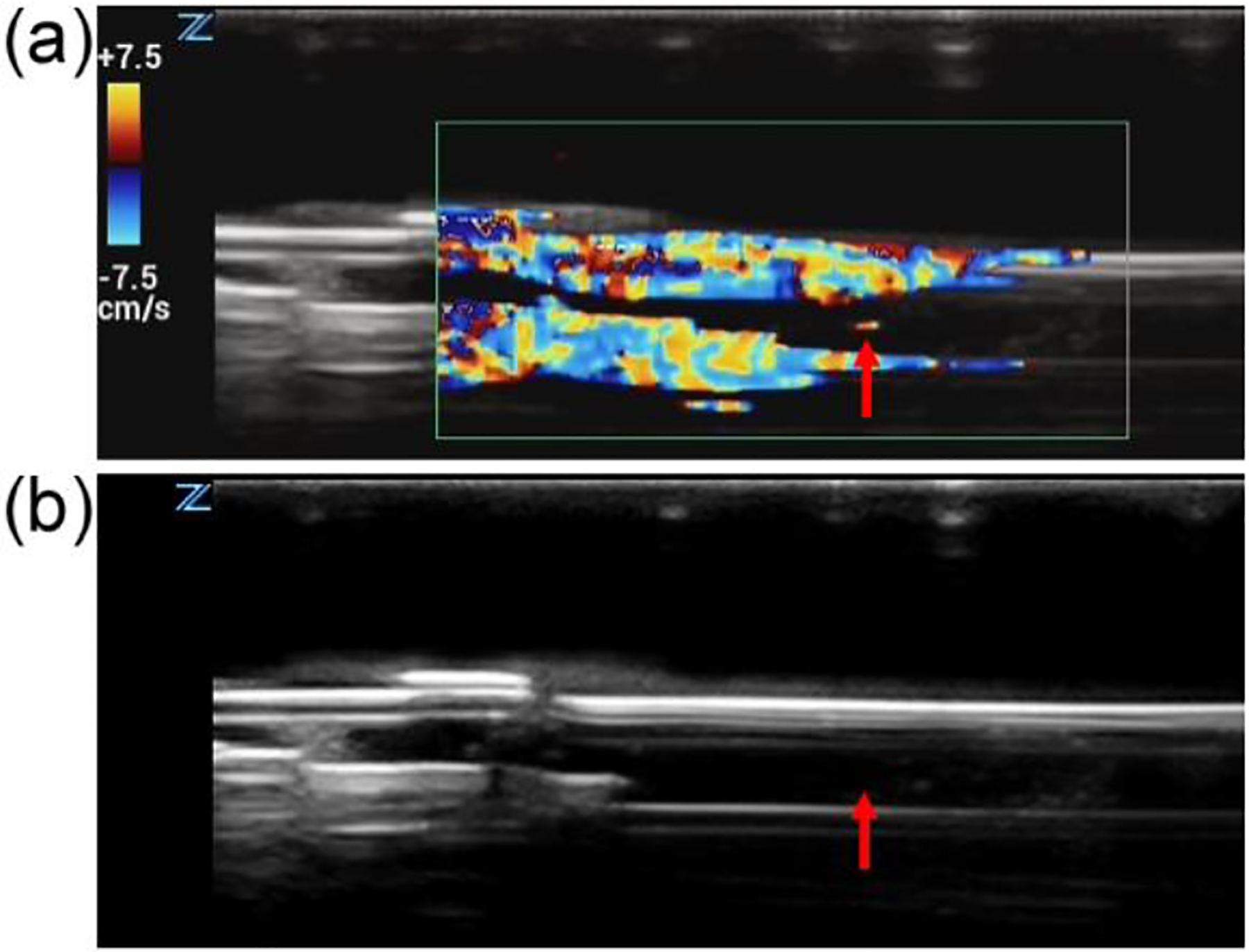
An in vitro blood flow-clot model was setup, and then an endovascular laser thrombolysis system was combined with high-intensity focused ultrasound to remove the clot. The laser and ultrasound pulses were synchronized and delivered to the blood clot concurrently. The laser pulses of 532 nm were delivered to the blood clot endovascularly through an optical fiber, whereas the ultrasound pulses of 0.5 MHz were applied noninvasively to the same region. Effectiveness of thrombolysis was evaluated by the ability to restore blood flow, which was monitored by ultrasound Doppler.
As laser powers increased, the ultrasound threshold pressures for effective thrombolysis decreased. For laser fluence levels of 0, 2, and 4 mJ/cm , the average negative ultrasound threshold pressures were 1.26 ± 0.114, 1.05 ± 0.181, and 0.59 ± 0.074 MPa, respectively. The periods of time needed to achieve effective thrombolysis were measured at 0.8, 2, and 4 mJ/cm laser fluence levels and 0.42, 0.70, and 0.98 MPa negative ultrasound pressures. In general, thrombolysis could be achieved more rapidly with higher laser powers or ultrasound pressures.
Effective thrombolysis can be achieved by combining endovascular laser with noninvasive ultrasound at relatively low power and pressure levels, which can potentially improve both the treatment efficiency and safety.
激光与超声的联合应用可通过增强空化效应显著提高溶栓效率。我们研发了一种基于光纤的激光-超声溶栓装置,并在体外血栓模型中测试了该技术恢复血流的可行性和效率。
建立了体外血流-血栓模型,然后将血管内激光溶栓系统与高强度聚焦超声联合使用以清除血栓。激光和超声脉冲同步发送至血栓部位,同时进行作用。532nm 的激光脉冲通过光纤经血管内输送至血栓部位,而 0.5MHz 的超声脉冲则经非侵入式施加于同一区域。通过超声多普勒监测血流恢复情况评估溶栓效果。
随着激光功率的增加,有效溶栓所需的超声阈值压力降低。对于激光能量密度分别为 0、2 和 4mJ/cm² 的情况,平均负超声阈值压力分别为 1.26±0.114、1.05±0.181 和 0.59±0.074MPa。在激光能量密度为 0.8、2 和 4mJ/cm²,负超声压力为 0.42、0.70 和 0.98MPa 的情况下,测量达到有效溶栓所需的时间。通常,随着激光功率或超声压力的增加,溶栓可以更快实现。
在相对较低的功率和压力水平下,通过将血管内激光与非侵入性超声相结合,可以实现有效的溶栓,这可能会提高治疗效率和安全性。