Jaykel Timothy J, Clark Michael S, Adamo Daniel A, Welch Brain T, Thompson Scott M, Young Jason R, Ehman Eric C
Department of Radiology, Mayo Clinic, Rochester, Minnesota, USA.
J Thorac Dis. 2020 Nov;12(11):6978-6991. doi: 10.21037/jtd-2019-cptn-09.
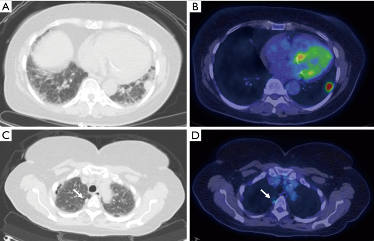
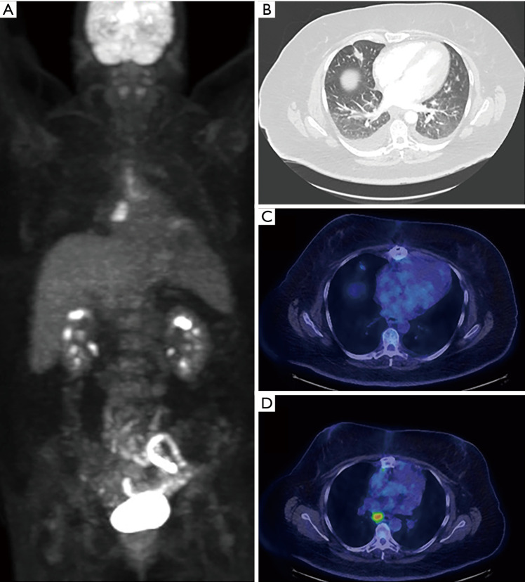
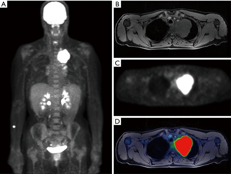
Ongoing technologic and therapeutic advancements in medicine are now testing the limits of conventional anatomic imaging techniques. The ability to image physiology, rather than simply anatomy, is critical in the management of multiple disease processes, especially in oncology. Nuclear medicine has assumed a leading role in detecting, diagnosing, staging and assessing treatment response of various pathologic entities, and appears well positioned to do so into the future. When combined with computed tomography (CT) or magnetic resonance imaging (MRI), positron emission tomography (PET) has become the technique of evaluating most solid tumors especially in the thorax. PET/CT serves as a key imaging modality in the initial evaluation of pulmonary nodules, often obviating the need for more invasive testing. PET/CT is essential to staging and restaging in bronchogenic carcinoma and offers key physiologic information with regard to treatment response. A more recent development, PET/MRI, shows promise in several specific lung cancer applications as well. Additional recent advancements in the field have allowed PET to expand beyond imaging with F-flurodeoxyglucose (FDG) alone, now with the ability to specifically image certain types of cell surface receptors. In the thorax this predominantly includes Ga-DOTATATE which targets the somatostatin receptors abundantly expressed in neuroendocrine tumors, including bronchial carcinoid. This receptor targeted imaging technique permits targeting these tumors with therapeutic analogues such as Lu labeled DOTATATE. Overall, the proper utilization of PET in the thorax has the ability to directly impact and improve patient care.
医学领域持续的技术和治疗进展正在考验传统解剖成像技术的极限。对生理功能而非仅仅解剖结构进行成像的能力在多种疾病过程的管理中至关重要,尤其是在肿瘤学领域。核医学在检测、诊断、分期和评估各种病理实体的治疗反应方面发挥了主导作用,并且在未来似乎也处于有利地位。当与计算机断层扫描(CT)或磁共振成像(MRI)相结合时,正电子发射断层扫描(PET)已成为评估大多数实体瘤,尤其是胸部实体瘤的技术。PET/CT是肺结节初始评估中的关键成像方式,常常无需进行更具侵入性的检查。PET/CT对于支气管肺癌的分期和再分期至关重要,并能提供有关治疗反应的关键生理信息。一项较新的进展,PET/MRI,在几种特定的肺癌应用中也显示出前景。该领域最近的其他进展使PET能够超越仅使用氟脱氧葡萄糖(FDG)成像,现在能够特异性地对某些类型的细胞表面受体进行成像。在胸部,这主要包括靶向生长抑素受体的镓[68Ga] DOTATATE,生长抑素受体在神经内分泌肿瘤(包括支气管类癌)中大量表达。这种受体靶向成像技术允许使用诸如镥[177Lu]标记的DOTATATE等治疗类似物来靶向这些肿瘤。总体而言,在胸部正确使用PET有能力直接影响并改善患者护理。