• 文献检索
  • 文档翻译
  • 深度研究
  • 学术资讯
  • Suppr Zotero 插件Zotero 插件
  • 邀请有礼
  • 套餐&价格
  • 历史记录
应用&插件
Suppr Zotero 插件Zotero 插件浏览器插件Mac 客户端Windows 客户端微信小程序
定价
高级版会员购买积分包购买API积分包
服务
文献检索文档翻译深度研究API 文档MCP 服务
关于我们
关于 Suppr公司介绍联系我们用户协议隐私条款
关注我们

Suppr 超能文献

核心技术专利:CN118964589B侵权必究
粤ICP备2023148730 号-1Suppr @ 2026

文献检索

告别复杂PubMed语法,用中文像聊天一样搜索,搜遍4000万医学文献。AI智能推荐,让科研检索更轻松。

立即免费搜索

文件翻译

保留排版,准确专业,支持PDF/Word/PPT等文件格式,支持 12+语言互译。

免费翻译文档

深度研究

AI帮你快速写综述,25分钟生成高质量综述,智能提取关键信息,辅助科研写作。

立即免费体验

MRI 预测 inverted papilloma 患者恶性转化:使用常规成像特征和表观扩散系数直方图分析的决策树分析。

MRI Predictors of Malignant Transformation in Patients with Inverted Papilloma: A Decision Tree Analysis Using Conventional Imaging Features and Histogram Analysis of Apparent Diffusion Coefficients.

机构信息

Department of Radiology and Research Institute of Radiology, University of Ulsan College of Medicine, Asan Medical Center, Seoul, Korea.

Department of Radiology, Chung-Ang University Hospital, Chung-Ang University College of Medicine, Seoul, Korea.

出版信息

Korean J Radiol. 2021 May;22(5):751-758. doi: 10.3348/kjr.2020.0576. Epub 2020 Nov 30.

DOI:10.3348/kjr.2020.0576
PMID:33289362
原文链接:https://pmc.ncbi.nlm.nih.gov/articles/PMC8076834/
Abstract

OBJECTIVE

Preoperative differentiation between inverted papilloma (IP) and its malignant transformation to squamous cell carcinoma (IP-SCC) is critical for patient management. We aimed to determine the diagnostic accuracy of conventional imaging features and histogram parameters obtained from whole tumor apparent diffusion coefficient (ADC) values to predict IP-SCC in patients with IP, using decision tree analysis.

MATERIALS AND METHODS

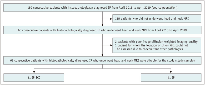
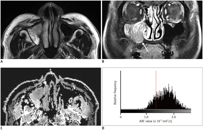
In this retrospective study, we analyzed data generated from the records of 180 consecutive patients with histopathologically diagnosed IP or IP-SCC who underwent head and neck magnetic resonance imaging, including diffusion-weighted imaging and 62 patients were included in the study. To obtain whole tumor ADC values, the region of interest was placed to cover the entire volume of the tumor. Classification and regression tree analyses were performed to determine the most significant predictors of IP-SCC among multiple covariates. The final tree was selected by cross-validation pruning based on minimal error.

RESULTS

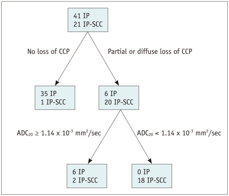
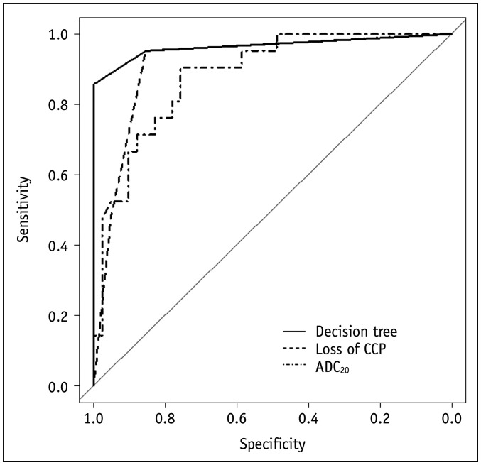
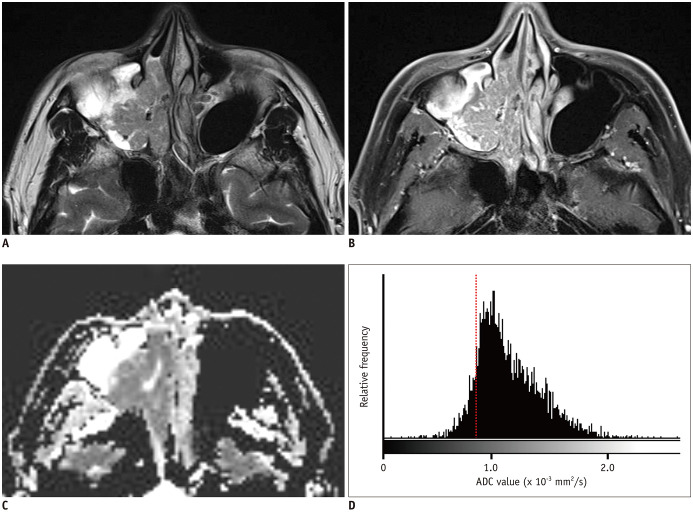
Of 62 patients with IP, 21 (34%) had IP-SCC. The decision tree analysis revealed that the loss of convoluted cerebriform pattern and the 20th percentile cutoff of ADC were the most significant predictors of IP-SCC. With these decision trees, the sensitivity, specificity, accuracy, and C-statistics were 86% (18 out of 21; 95% confidence interval [CI], 65-95%), 100% (41 out of 41; 95% CI, 91-100%), 95% (59 out of 61; 95% CI, 87-98%), and 0.966 (95% CI, 0.912-1.000), respectively.

CONCLUSION

Decision tree analysis using conventional imaging features and histogram analysis of whole volume ADC could predict IP-SCC in patients with IP with high diagnostic accuracy.

摘要

目的

术前鉴别内翻性乳头状瘤(IP)及其恶性转化为鳞状细胞癌(IP-SCC)对于患者管理至关重要。我们旨在通过决策树分析确定常规成像特征和直方图参数与全肿瘤表观扩散系数(ADC)值之间的诊断准确性,以预测 IP 患者中的 IP-SCC。

材料与方法

在这项回顾性研究中,我们分析了 180 例经组织病理学诊断为 IP 或 IP-SCC 的连续患者的头颈部磁共振成像(包括扩散加权成像)记录数据,其中 62 例患者纳入研究。为了获得全肿瘤 ADC 值,感兴趣区域被放置以覆盖肿瘤的整个体积。分类和回归树分析用于确定多个协变量中 IP-SCC 的最显著预测因子。最终树是通过基于最小误差的交叉验证修剪选择的。

结果

在 62 例 IP 患者中,21 例(34%)患有 IP-SCC。决策树分析显示,脑回状模式的丧失和 ADC 的第 20 百分位截断是 IP-SCC 的最显著预测因子。使用这些决策树,敏感性、特异性、准确性和 C 统计量分别为 86%(21 例中的 18 例;95%置信区间 [CI],65-95%)、100%(41 例中的 41 例;95% CI,91-100%)、95%(61 例中的 59 例;95% CI,87-98%)和 0.966(95% CI,0.912-1.000)。

结论

使用常规成像特征和全容积 ADC 直方图分析的决策树分析可以预测 IP 患者中的 IP-SCC,具有较高的诊断准确性。

https://cdn.ncbi.nlm.nih.gov/pmc/blobs/4295/8076834/14ac0b33d9ae/kjr-22-751-g005.jpg
https://cdn.ncbi.nlm.nih.gov/pmc/blobs/4295/8076834/c7deffbf964b/kjr-22-751-g001.jpg
https://cdn.ncbi.nlm.nih.gov/pmc/blobs/4295/8076834/89854ff3b44e/kjr-22-751-g002.jpg
https://cdn.ncbi.nlm.nih.gov/pmc/blobs/4295/8076834/1862b7b922e6/kjr-22-751-g003.jpg
https://cdn.ncbi.nlm.nih.gov/pmc/blobs/4295/8076834/8e3c37ea3965/kjr-22-751-g004.jpg
https://cdn.ncbi.nlm.nih.gov/pmc/blobs/4295/8076834/14ac0b33d9ae/kjr-22-751-g005.jpg

相似文献

1
MRI Predictors of Malignant Transformation in Patients with Inverted Papilloma: A Decision Tree Analysis Using Conventional Imaging Features and Histogram Analysis of Apparent Diffusion Coefficients.MRI 预测 inverted papilloma 患者恶性转化:使用常规成像特征和表观扩散系数直方图分析的决策树分析。
Korean J Radiol. 2021 May;22(5):751-758. doi: 10.3348/kjr.2020.0576. Epub 2020 Nov 30.
2
Differentiation of squamous cell carcinoma and inverted papilloma using non-invasive MR perfusion imaging.使用非侵入性磁共振灌注成像鉴别鳞状细胞癌和内翻性乳头状瘤
Dentomaxillofac Radiol. 2015;44(9):20150074. doi: 10.1259/dmfr.20150074. Epub 2015 Jun 9.
3
Imaging predictors for malignant transformation of inverted papilloma.内翻性乳头状瘤恶变的影像学预测指标
Laryngoscope. 2019 Apr;129(4):777-782. doi: 10.1002/lary.27582. Epub 2018 Dec 4.
4
Clinical evidence based review and systematic scientific review in the identification of malignant transformation of inverted papilloma.临床循证综述和系统科学综述在确定内翻性乳头状瘤恶变中的作用。
J Otolaryngol Head Neck Surg. 2020 Apr 30;49(1):25. doi: 10.1186/s40463-020-00420-x.
5
Development and validation of apparent diffusion coefficient histogram-based nomogram for predicting malignant transformation of sinonasal inverted papilloma.基于表观扩散系数直方图的nomogram 模型预测鼻窦内翻性乳头状瘤恶变的建立与验证。
Dentomaxillofac Radiol. 2023 Sep;52(6):20220301. doi: 10.1259/dmfr.20220301. Epub 2023 Feb 17.
6
Multiparametric MRI-based radiomics nomogram for predicting malignant transformation of sinonasal inverted papilloma.基于多参数磁共振成像的影像组学列线图预测鼻窦内翻性乳头状瘤的恶变
Clin Radiol. 2024 Mar;79(3):e408-e416. doi: 10.1016/j.crad.2023.11.004. Epub 2023 Nov 22.
7
Histogram analysis of apparent diffusion coefficient maps for the differentiation between lymphoma and metastatic lymph nodes of squamous cell carcinoma in head and neck region.头颈部区域淋巴瘤与鳞状细胞癌转移性淋巴结鉴别的表观扩散系数图直方图分析
Acta Radiol. 2018 Jun;59(6):672-680. doi: 10.1177/0284185117730688. Epub 2017 Sep 4.
8
Microvessel barrier dysfunction in sinonasal inverted papilloma-associated squamous cell carcinoma and its manifestation in dynamic contrast-enhanced MRI.鼻窦内翻性乳头状瘤相关鳞状细胞癌微血管屏障功能障碍及其在动态对比增强 MRI 中的表现。
Int Forum Allergy Rhinol. 2024 Jul;14(7):1173-1181. doi: 10.1002/alr.23316. Epub 2024 Jan 21.
9
Clinical management of squamous cell carcinoma associated with sinonasal inverted papilloma.鼻窦内翻性乳头状瘤相关鳞状细胞癌的临床管理
Auris Nasus Larynx. 2017 Feb;44(1):98-103. doi: 10.1016/j.anl.2016.04.004. Epub 2016 Jun 1.
10
Predictive Value of Magnetic Resonance Imaging Multi-parametric Analysis for Malignant Transformation of Sinonasal Inverted Papilloma: A Comprehensive Prediction Model.磁共振成像多参数分析对鼻窦内翻性乳头状瘤恶变的预测价值:综合预测模型。
Curr Med Imaging. 2023;19(6):596-604. doi: 10.2174/1573405618666220928091936.

引用本文的文献

1
Identifying sinonasal inverted papilloma by machine learning: a systematic review and meta-analysis.通过机器学习识别鼻窦内翻性乳头状瘤:一项系统评价和荟萃分析。
Front Oncol. 2025 Aug 26;15:1628999. doi: 10.3389/fonc.2025.1628999. eCollection 2025.
2
Assessing MRI interpretability of the orbit, paranasal sinuses, and nasopharynx in cochlear implant patients.评估人工耳蜗植入患者眼眶、鼻窦和鼻咽部的MRI可解释性。
Front Neurol. 2025 Jul 31;16:1636128. doi: 10.3389/fneur.2025.1636128. eCollection 2025.
3
Differences in Imaging and Histology Between Sinonasal Inverted Papilloma with and Without Squamous Cell Carcinoma.

本文引用的文献

1
Application of diffusion-weighted MR imaging with ADC measurement for distinguishing between the histopathological types of sinonasal neoplasms.扩散加权磁共振成像 ADC 值测量在鉴别鼻窦肿瘤组织学类型中的应用。
Clin Imaging. 2019 May-Jun;55:76-82. doi: 10.1016/j.clinimag.2019.02.004. Epub 2019 Feb 7.
2
Imaging predictors for malignant transformation of inverted papilloma.内翻性乳头状瘤恶变的影像学预测指标
Laryngoscope. 2019 Apr;129(4):777-782. doi: 10.1002/lary.27582. Epub 2018 Dec 4.
3
Can Diffusion Weighted Imaging Aid in Differentiating Benign from Malignant Sinonasal Masses?: A Useful Adjunct.
伴与不伴鳞状细胞癌的鼻窦内翻性乳头状瘤在影像学和组织学上的差异
Diagnostics (Basel). 2025 Jun 27;15(13):1645. doi: 10.3390/diagnostics15131645.
4
Preoperative prediction of malignant transformation in sinonasal inverted papilloma: a novel MRI-based deep learning approach.鼻腔鼻窦内翻性乳头状瘤恶变的术前预测:一种基于MRI的新型深度学习方法。
Eur Radiol. 2025 May 12. doi: 10.1007/s00330-025-11655-5.
5
Safety and efficacy of selumetinib in pediatric and adult patients with neurofibromatosis type 1 and plexiform neurofibroma.司美替尼在1型神经纤维瘤病和丛状神经纤维瘤儿科及成年患者中的安全性和有效性。
Neuro Oncol. 2024 Dec 5;26(12):2352-2363. doi: 10.1093/neuonc/noae121.
6
[Inverted papilloma of the nose and paranasal sinuses : Diagnosis, treatment, and malignant transformation].[鼻及鼻窦内翻性乳头状瘤:诊断、治疗及恶变]
HNO. 2024 Apr;72(4):257-264. doi: 10.1007/s00106-023-01406-7. Epub 2024 Jan 12.
7
Towards in vivo characterization of thyroid nodules suspicious for malignancy using multispectral optoacoustic tomography.使用多光谱光声断层成像术对疑似恶性甲状腺结节进行体内特征分析。
Eur J Nucl Med Mol Imaging. 2023 Jul;50(9):2736-2750. doi: 10.1007/s00259-023-06189-1. Epub 2023 Apr 11.
8
A new biomarker combining multimodal MRI radiomics and clinical indicators for differentiating inverted papilloma from nasal polyp invaded the olfactory nerve possibly.一种结合多模态MRI影像组学和临床指标的新型生物标志物,可能用于鉴别内翻性乳头状瘤与可能侵犯嗅神经的鼻息肉。
Front Neurol. 2023 Mar 21;14:1151455. doi: 10.3389/fneur.2023.1151455. eCollection 2023.
9
Development and validation of apparent diffusion coefficient histogram-based nomogram for predicting malignant transformation of sinonasal inverted papilloma.基于表观扩散系数直方图的nomogram 模型预测鼻窦内翻性乳头状瘤恶变的建立与验证。
Dentomaxillofac Radiol. 2023 Sep;52(6):20220301. doi: 10.1259/dmfr.20220301. Epub 2023 Feb 17.
10
Preoperative Prediction of Malignant Transformation of Sinonasal Inverted Papilloma Using MR Radiomics.利用磁共振成像放射组学对鼻窦内翻性乳头状瘤恶变进行术前预测
Front Oncol. 2022 Mar 23;12:870544. doi: 10.3389/fonc.2022.870544. eCollection 2022.
扩散加权成像能否有助于鉴别鼻窦良恶性肿物?:一种有用的辅助手段。
Pol J Radiol. 2017 Jun 28;82:345-355. doi: 10.12659/PJR.900633. eCollection 2017.
4
MRI-Based Texture Analysis to Differentiate Sinonasal Squamous Cell Carcinoma from Inverted Papilloma.基于磁共振成像的纹理分析鉴别鼻窦鳞状细胞癌与内翻性乳头状瘤
AJNR Am J Neuroradiol. 2017 May;38(5):1019-1025. doi: 10.3174/ajnr.A5106. Epub 2017 Mar 2.
5
STARD 2015: An Updated List of Essential Items for Reporting Diagnostic Accuracy Studies.STARD 2015:报告诊断准确性研究的基本项目的更新清单。
Radiology. 2015 Dec;277(3):826-32. doi: 10.1148/radiol.2015151516. Epub 2015 Oct 28.
6
Histogram Analysis of Apparent Diffusion Coefficients for Occult Tonsil Cancer in Patients with Cervical Nodal Metastasis from an Unknown Primary Site at Presentation.基于 MRI 的表观扩散系数直方图分析对原发灶不明颈部转移鳞癌隐匿性扁桃体癌的诊断价值
Radiology. 2016 Jan;278(1):146-55. doi: 10.1148/radiol.2015141727. Epub 2015 Jun 24.
7
Differentiation of squamous cell carcinoma and inverted papilloma using non-invasive MR perfusion imaging.使用非侵入性磁共振灌注成像鉴别鳞状细胞癌和内翻性乳头状瘤
Dentomaxillofac Radiol. 2015;44(9):20150074. doi: 10.1259/dmfr.20150074. Epub 2015 Jun 9.
8
Malignant transformation of sinonasal inverted papilloma: A retrospective analysis of 32 cases.鼻窦内翻性乳头状瘤的恶变:32例回顾性分析
Oncol Lett. 2014 Dec;8(6):2637-2641. doi: 10.3892/ol.2014.2539. Epub 2014 Sep 16.
9
18F-FDG PET/CT findings of sinonasal inverted papilloma with or without coexistent malignancy: comparison with MR imaging findings in eight patients.伴或不伴并存恶性肿瘤的鼻窦内翻性乳头状瘤的18F-FDG PET/CT表现:与8例患者的磁共振成像表现对比
Neuroradiology. 2009 Apr;51(4):265-71. doi: 10.1007/s00234-009-0510-2. Epub 2009 Mar 4.
10
Sinonasal inverted papilloma: value of convoluted cerebriform pattern on MR imaging.鼻窦内翻性乳头状瘤:磁共振成像上脑回样形态的价值
AJNR Am J Neuroradiol. 2008 Sep;29(8):1556-60. doi: 10.3174/ajnr.A1128. Epub 2008 May 22.