• 文献检索
  • 文档翻译
  • 深度研究
  • 学术资讯
  • Suppr Zotero 插件Zotero 插件
  • 邀请有礼
  • 套餐&价格
  • 历史记录
应用&插件
Suppr Zotero 插件Zotero 插件浏览器插件Mac 客户端Windows 客户端微信小程序
定价
高级版会员购买积分包购买API积分包
服务
文献检索文档翻译深度研究API 文档MCP 服务
关于我们
关于 Suppr公司介绍联系我们用户协议隐私条款
关注我们

Suppr 超能文献

核心技术专利:CN118964589B侵权必究
粤ICP备2023148730 号-1Suppr @ 2026

文献检索

告别复杂PubMed语法,用中文像聊天一样搜索,搜遍4000万医学文献。AI智能推荐,让科研检索更轻松。

立即免费搜索

文件翻译

保留排版,准确专业,支持PDF/Word/PPT等文件格式,支持 12+语言互译。

免费翻译文档

深度研究

AI帮你快速写综述,25分钟生成高质量综述,智能提取关键信息,辅助科研写作。

立即免费体验

利用多模态成像技术研究糖尿病视网膜病变中黄斑灌注状态与直至远周边部的微血管参数之间的关联。

Association of macular perfusion status with microvascular parameters up to the far periphery in diabetic retinopathy using multimodal imaging.

作者信息

Hajdu Dorottya, Sedova Aleksandra, Datlinger Felix, Hafner Julia, Steiner Irene, Kriechbaum Katharina, Scholda Christoph, Sacu Stefan, Schmidt-Erfurth Ursula, Pollreisz Andreas

机构信息

Department of Ophthalmology and Optometry, Vienna Clinical Trial Centre (VTC), Medical University of Vienna, Waehringer Guertel 18-20, E8i, 1090, Vienna, Austria.

Center for Medical Statistics, Informatics, and Intelligent Systems (CeMSIIS), Section for Medical Statistics, Medical University of Vienna, Vienna, Austria.

出版信息

Int J Retina Vitreous. 2020 Nov 4;6(1):50. doi: 10.1186/s40942-020-00253-w.

DOI:10.1186/s40942-020-00253-w
PMID:33292856
原文链接:https://pmc.ncbi.nlm.nih.gov/articles/PMC7640640/
Abstract

BACKGROUND

The aim of our study was to investigate a possible association between macular perfusion status and retinal ischemia and leakage up to far peripheral retinal areas in eyes with early to advanced stages of diabetic retinopathy (DR).

METHODS

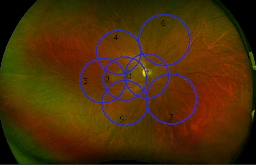
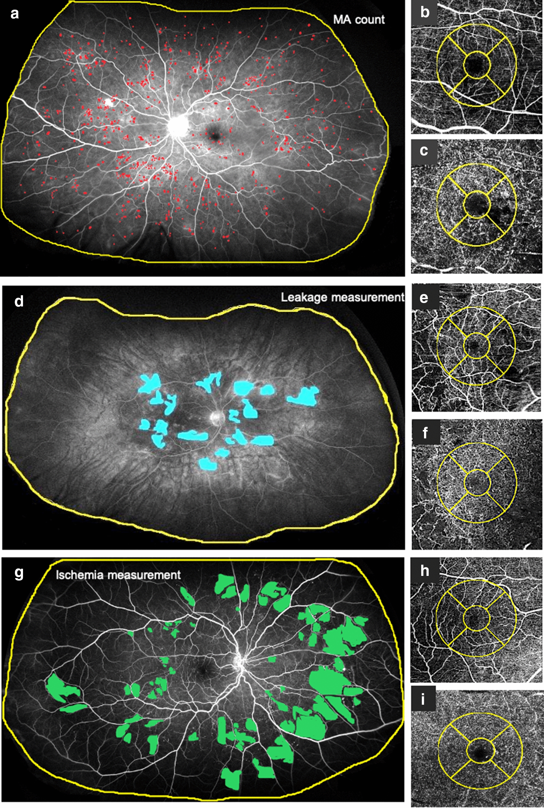
In a retrospective, cross sectional analysis ultrawide field (UWF) color fundus photos (Optos, Optomap California) were graded for DR severity. Foveal avascular zone (FAZ) and vessel density from the superficial (SCP) and deep capillary plexus (DCP) were assessed on optical coherence tomography angiography (OCTA) scans (Topcon, DRI-OCT Triton). UWF angiography images were used to quantify leakage/ischemic index and number of microaneurysms (MA). Age, gender, disease duration, type of diabetes, HbA1C, hypertension, complications of diabetes and ocular history were recorded. Univariate mixed models and Spearman correlation analysis were used for statistical testing.

RESULTS

24 eyes of 17 laser-naive diabetic patients with different stages of DR were analyzed. The mean age was 59.56 ± 8.46 years and the mean disease duration 19.65 ± 12.25 years. No statistically significant associations between FAZ size, macular vessel density of SCP/DCP and peripheral retinal ischemia, leakage and MA number were demonstrated. Higher stages of DR were associated with ischemic index (estimate [95% CI]: 13.04 [1.5; 24.5], p = 0.033) and MA count (estimate [95% CI]: 43.7 [15.6; 71.8], p = 0.01), but no association with leakage index was observed. Only weak correlations between DR severity and anamnestic data were found.

CONCLUSION

Retinal ischemic index and the amount of MAs assessed on UWFA up to peripheral areas are indicators of DR severity but not related to microvascular perfusion status in the macular region. Significance and timely sequence of macular vessel density in DR progression may need to be re-evaluated in future studies.

摘要

背景

我们研究的目的是调查糖尿病视网膜病变(DR)早期至晚期患者黄斑灌注状态与视网膜缺血及远周边视网膜区域渗漏之间可能存在的关联。

方法

在一项回顾性横断面分析中,对超广角(UWF)彩色眼底照片(Optos,Optomap California)进行DR严重程度分级。在光学相干断层扫描血管造影(OCTA)扫描(Topcon,DRI - OCT Triton)上评估中央凹无血管区(FAZ)以及浅层(SCP)和深层毛细血管丛(DCP)的血管密度。使用UWF血管造影图像量化渗漏/缺血指数和微动脉瘤(MA)数量。记录年龄、性别、病程、糖尿病类型、糖化血红蛋白、高血压、糖尿病并发症和眼部病史。采用单变量混合模型和Spearman相关性分析进行统计学检验。

结果

分析了17例未接受激光治疗的不同阶段DR糖尿病患者的24只眼。平均年龄为59.56±8.46岁,平均病程为19.65±12.25年。未发现FAZ大小、SCP/DCP黄斑血管密度与周边视网膜缺血、渗漏及MA数量之间存在统计学显著关联。DR更高阶段与缺血指数(估计值[95%置信区间]:13.04[1.5;24.5],p = 0.033)和MA计数(估计值[95%置信区间]:43.7[15.6;71.8],p = 0.01)相关,但未观察到与渗漏指数相关。仅发现DR严重程度与既往病史数据之间存在弱相关性。

结论

在外周区域通过超广角荧光血管造影(UWFA)评估的视网膜缺血指数和MA数量是DR严重程度的指标,但与黄斑区域的微血管灌注状态无关。黄斑血管密度在DR进展中的意义和时间顺序可能需要在未来研究中重新评估。

https://cdn.ncbi.nlm.nih.gov/pmc/blobs/f17c/7640640/632683640bb1/40942_2020_253_Fig2_HTML.jpg
https://cdn.ncbi.nlm.nih.gov/pmc/blobs/f17c/7640640/34d54e05eb93/40942_2020_253_Fig1_HTML.jpg
https://cdn.ncbi.nlm.nih.gov/pmc/blobs/f17c/7640640/632683640bb1/40942_2020_253_Fig2_HTML.jpg
https://cdn.ncbi.nlm.nih.gov/pmc/blobs/f17c/7640640/34d54e05eb93/40942_2020_253_Fig1_HTML.jpg
https://cdn.ncbi.nlm.nih.gov/pmc/blobs/f17c/7640640/632683640bb1/40942_2020_253_Fig2_HTML.jpg

相似文献

1
Association of macular perfusion status with microvascular parameters up to the far periphery in diabetic retinopathy using multimodal imaging.利用多模态成像技术研究糖尿病视网膜病变中黄斑灌注状态与直至远周边部的微血管参数之间的关联。
Int J Retina Vitreous. 2020 Nov 4;6(1):50. doi: 10.1186/s40942-020-00253-w.
2
Macular Perfusion Deficits on OCT Angiography Correlate with Nonperfusion on Ultrawide-field Fluorescein Angiography in Diabetic Retinopathy.OCT 血管造影上的黄斑灌注缺损与糖尿病视网膜病变的超广角荧光素血管造影上的无灌注相关。
Ophthalmol Retina. 2023 Aug;7(8):692-702. doi: 10.1016/j.oret.2023.04.003. Epub 2023 Apr 14.
3
Identification of microvascular and morphological alterations in eyes with central retinal non-perfusion.中央视网膜无灌注眼中小血管和形态改变的鉴定。
PLoS One. 2020 Nov 10;15(11):e0241753. doi: 10.1371/journal.pone.0241753. eCollection 2020.
4
Macular Capillary Perfusion in Chinese Patients With Diabetic Retinopathy Obtained With Optical Coherence Tomography Angiography.光学相干断层扫描血管造影术检测中国糖尿病视网膜病变患者的黄斑区毛细血管灌注情况
Ophthalmic Surg Lasers Imaging Retina. 2019 Apr 1;50(4):e88-e95. doi: 10.3928/23258160-20190401-12.
5
Combined Multimodal Analysis of Peripheral Retinal and Macular Circulation in Diabetic Retinopathy (COPRA Study).糖尿病视网膜病变外周视网膜和黄斑循环的联合多模态分析(COPRA研究)
Ophthalmol Retina. 2019 Jul;3(7):580-588. doi: 10.1016/j.oret.2019.03.001. Epub 2019 Mar 20.
6
Quantitative Parameters from OCT Angiography in Patients with Diabetic Retinopathy and in Those with Only Peripheral Retinopathy Compared with Control Participants.糖尿病视网膜病变患者、仅患有周边视网膜病变患者与对照参与者相比,光学相干断层扫描血管造影的定量参数。
Ophthalmol Sci. 2021 May 26;1(2):100030. doi: 10.1016/j.xops.2021.100030. eCollection 2021 Jun.
7
Correlation Analysis between Foveal Avascular Zone and Peripheral Ischemic Index in Diabetic Retinopathy: A Pilot Study.糖尿病视网膜病变中黄斑无血管区与周边缺血指数的相关性分析:一项初步研究
Ophthalmol Retina. 2018 Jan;2(1):46-52. doi: 10.1016/j.oret.2017.05.007. Epub 2017 Jul 26.
8
Optical coherence tomography angiography discerns preclinical diabetic retinopathy in eyes of patients with type 2 diabetes without clinical diabetic retinopathy.光学相干断层扫描血管造影术可在无临床糖尿病视网膜病变的 2 型糖尿病患者眼中识别出临床前期糖尿病性视网膜病变。
Acta Diabetol. 2018 May;55(5):469-477. doi: 10.1007/s00592-018-1115-1. Epub 2018 Feb 16.
9
Multimodal Retinal Imaging in Patients with Diabetes Mellitus and Association with Cerebrovascular Disease.糖尿病患者的多模态视网膜成像及其与脑血管疾病的关联。
Ophthalmic Res. 2023;66(1):1044-1052. doi: 10.1159/000531249. Epub 2023 May 30.
10
Macular vessels density in diabetic retinopathy: quantitative assessment using optical coherence tomography angiography.糖尿病视网膜病变中的黄斑血管密度:使用光学相干断层扫描血管造影术进行定量评估。
Int Ophthalmol. 2019 Aug;39(8):1845-1859. doi: 10.1007/s10792-018-1013-0. Epub 2018 Sep 7.

引用本文的文献

1
Predictors of Peripheral Retinal Non-Perfusion in Clinically Significant Diabetic Macular Edema.临床显著性糖尿病黄斑水肿患者周边视网膜无灌注的预测因素
J Clin Med. 2024 Dec 26;14(1):52. doi: 10.3390/jcm14010052.
2
Imaging Modalities for Assessing the Vascular Component of Diabetic Retinal Disease: Review and Consensus for an Updated Staging System.评估糖尿病视网膜病变血管成分的成像方式:更新分期系统的综述与共识
Ophthalmol Sci. 2023 Dec 10;4(3):100449. doi: 10.1016/j.xops.2023.100449. eCollection 2024 May-Jun.
3
Relationship between nonperfusion area from widefield optical coherence tomography angiography and macular vascular parameters in diabetic retinopathy.

本文引用的文献

1
Retinal Capillary Plexus Pattern and Density from Fovea to Periphery Measured in Healthy Eyes with Swept-Source Optical Coherence Tomography Angiography.应用扫频源光学相干断层血管成像术测量健康人眼从中心凹至周边的视网膜毛细血管丛形态和密度。
Sci Rep. 2020 Jan 30;10(1):1474. doi: 10.1038/s41598-020-58359-y.
2
Changes in retinal and choriocapillaris density in diabetic patients receiving anti-vascular endothelial growth factor treatment using optical coherence tomography angiography.使用光学相干断层扫描血管造影术对接受抗血管内皮生长因子治疗的糖尿病患者视网膜和脉络膜毛细血管密度的变化
Int J Retina Vitreous. 2019 Dec 10;5:41. doi: 10.1186/s40942-019-0192-9. eCollection 2019.
3
广角光相干断层扫描血管造影的无灌注区与糖尿病视网膜病变的黄斑血管参数的关系。
Int Ophthalmol. 2023 Dec;43(12):4803-4814. doi: 10.1007/s10792-023-02882-0. Epub 2023 Oct 18.
4
[Diagnosis, treatment and monitoring of diabetic eye disease (Update 2023)].糖尿病眼病的诊断、治疗及监测(2023年更新)
Wien Klin Wochenschr. 2023 Jan;135(Suppl 1):195-200. doi: 10.1007/s00508-022-02119-7. Epub 2023 Apr 20.
5
Macular Perfusion Deficits on OCT Angiography Correlate with Nonperfusion on Ultrawide-field Fluorescein Angiography in Diabetic Retinopathy.OCT 血管造影上的黄斑灌注缺损与糖尿病视网膜病变的超广角荧光素血管造影上的无灌注相关。
Ophthalmol Retina. 2023 Aug;7(8):692-702. doi: 10.1016/j.oret.2023.04.003. Epub 2023 Apr 14.
6
A lightweight deep learning model for automatic segmentation and analysis of ophthalmic images.一种用于眼科图像自动分割和分析的轻量级深度学习模型。
Sci Rep. 2022 May 20;12(1):8508. doi: 10.1038/s41598-022-12486-w.
7
1-Year Fixed-Regimen Bevacizumab Treatment in DME-Vascular Network Image Analysis in Optical Coherence Tomography Angiography Study.光学相干断层扫描血管造影研究中糖尿病性黄斑水肿-血管网络图像分析的1年固定方案贝伐单抗治疗
J Clin Med. 2022 Apr 11;11(8):2125. doi: 10.3390/jcm11082125.
8
Intraprocedural OCT monitoring of the immediate treatment response during indocyanine green angiography-guided laser therapy of teleangiectatic capillaries in diabetic macular edema.术中 OCT 监测吲哚菁绿引导的激光治疗糖尿病黄斑水肿毛细血管扩张症即刻治疗反应。
Sci Rep. 2022 Feb 10;12(1):2315. doi: 10.1038/s41598-022-05950-0.
9
Featureless retina in diabetic retinopathy: Clinical and fluorescein angiographic profile.糖尿病性视网膜病变无特征性视网膜:临床和荧光素血管造影特征。
Indian J Ophthalmol. 2021 Nov;69(11):3194-3198. doi: 10.4103/ijo.IJO_1352_21.
PRESENCE OF PERIPHERAL LESIONS AND CORRELATION TO MACULAR PERFUSION, OXYGENATION AND NEURODEGENERATION IN EARLY TYPE II DIABETIC RETINAL DISEASE.
早期 II 型糖尿病视网膜病变中外周病变的存在及其与黄斑灌注、氧合和神经退行性变的相关性。
Retina. 2020 Oct;40(10):1964-1971. doi: 10.1097/IAE.0000000000002704.
4
Widefield OCT-Angiography and Fluorescein Angiography Assessments of Nonperfusion in Diabetic Retinopathy and Edema Treated with Anti-Vascular Endothelial Growth Factor.宽视野 OCT 血管造影和荧光素血管造影评估抗血管内皮生长因子治疗糖尿病视网膜病变和水肿的无灌注区。
Ophthalmology. 2019 Dec;126(12):1685-1694. doi: 10.1016/j.ophtha.2019.06.022. Epub 2019 Jun 26.
5
Quantitative Ultra-Widefield Angiography and Diabetic Retinopathy Severity: An Assessment of Panretinal Leakage Index, Ischemic Index and Microaneurysm Count.定量超广角血管造影与糖尿病视网膜病变严重程度:对全视网膜渗漏指数、缺血指数和微动脉瘤计数的评估。
Ophthalmology. 2019 Nov;126(11):1527-1532. doi: 10.1016/j.ophtha.2019.05.034. Epub 2019 Jun 8.
6
Classification and Guidelines for Widefield Imaging: Recommendations from the International Widefield Imaging Study Group.宽视野成像的分类与指南:国际宽视野成像研究组的建议
Ophthalmol Retina. 2019 Oct;3(10):843-849. doi: 10.1016/j.oret.2019.05.007. Epub 2019 May 13.
7
Optical coherence tomography angiography analysis of macular vessel density before and after anti-VEGF therapy in eyes with diabetic retinopathy.糖尿病视网膜病变患者抗VEGF治疗前后黄斑血管密度的光学相干断层扫描血管造影分析
Int Ophthalmol. 2019 Oct;39(10):2361-2371. doi: 10.1007/s10792-019-01076-x. Epub 2019 May 22.
8
Combined Multimodal Analysis of Peripheral Retinal and Macular Circulation in Diabetic Retinopathy (COPRA Study).糖尿病视网膜病变外周视网膜和黄斑循环的联合多模态分析(COPRA研究)
Ophthalmol Retina. 2019 Jul;3(7):580-588. doi: 10.1016/j.oret.2019.03.001. Epub 2019 Mar 20.
9
Correlation Analysis between Foveal Avascular Zone and Peripheral Ischemic Index in Diabetic Retinopathy: A Pilot Study.糖尿病视网膜病变中黄斑无血管区与周边缺血指数的相关性分析:一项初步研究
Ophthalmol Retina. 2018 Jan;2(1):46-52. doi: 10.1016/j.oret.2017.05.007. Epub 2017 Jul 26.
10
Global prevalence of diabetic retinopathy: protocol for a systematic review and meta-analysis.全球糖尿病视网膜病变患病率的系统评价和 Meta 分析方案。
BMJ Open. 2019 Mar 3;9(3):e022188. doi: 10.1136/bmjopen-2018-022188.