Department of Sleep and Cognition, Netherlands Institute for Neuroscience, an Institute of the Royal Netherlands Academy of Arts and Sciences, Amsterdam, The Netherlands.
Department of Integrative Neurophysiology, Center for Neurogenomics and Cognitive Research (CNCR), Amsterdam Neuroscience, VU University Amsterdam, Amsterdam, The Netherlands.
Transl Psychiatry. 2020 Dec 8;10(1):425. doi: 10.1038/s41398-020-01109-5.
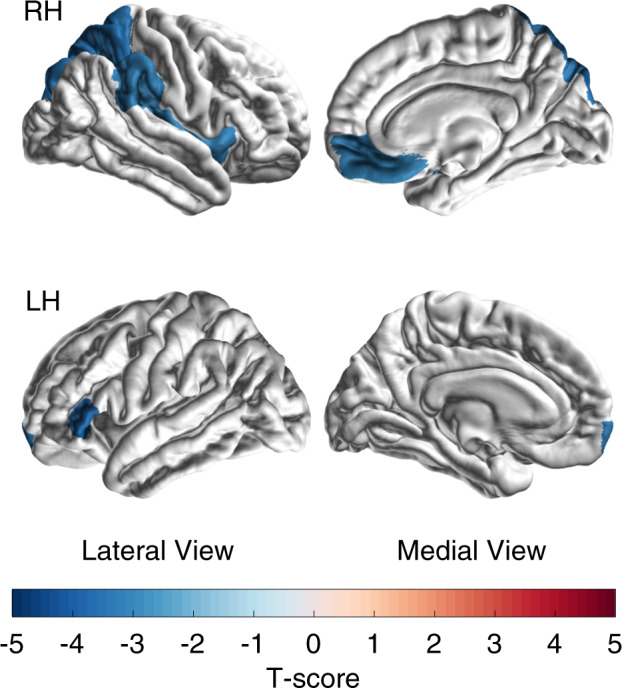
It has been difficult to find robust brain structural correlates of the overall severity of major depressive disorder (MDD). We hypothesized that specific symptoms may better reveal correlates and investigated this for the severity of insomnia, both a key symptom and a modifiable major risk factor of MDD. Cortical thickness, surface area and subcortical volumes were assessed from T1-weighted brain magnetic resonance imaging (MRI) scans of 1053 MDD patients (age range 13-79 years) from 15 cohorts within the ENIGMA MDD Working Group. Insomnia severity was measured by summing the insomnia items of the Hamilton Depression Rating Scale (HDRS). Symptom specificity was evaluated with correlates of overall depression severity. Disease specificity was evaluated in two independent samples comprising 2108 healthy controls, and in 260 clinical controls with bipolar disorder. Results showed that MDD patients with more severe insomnia had a smaller cortical surface area, mostly driven by the right insula, left inferior frontal gyrus pars triangularis, left frontal pole, right superior parietal cortex, right medial orbitofrontal cortex, and right supramarginal gyrus. Associations were specific for insomnia severity, and were not found for overall depression severity. Associations were also specific to MDD; healthy controls and clinical controls showed differential insomnia severity association profiles. The findings indicate that MDD patients with more severe insomnia show smaller surfaces in several frontoparietal cortical areas. While explained variance remains small, symptom-specific associations could bring us closer to clues on underlying biological phenomena of MDD.
一直难以找到重度抑郁症(MDD)整体严重程度的稳健大脑结构相关性。我们假设特定症状可能更好地揭示相关性,并针对失眠的严重程度进行了研究,失眠既是 MDD 的主要症状,也是可改变的主要危险因素之一。从 15 个 ENIGMA MDD 工作组队列中的 1053 名 MDD 患者(年龄范围为 13-79 岁)的 T1 加权脑磁共振成像(MRI)扫描中评估了皮质厚度、表面积和皮质下体积。失眠严重程度通过汉密尔顿抑郁评定量表(HDRS)的失眠项目总和来衡量。使用与总体抑郁严重程度相关的指标来评估症状特异性。在两个独立的样本中评估疾病特异性,包括 2108 名健康对照者和 260 名双相情感障碍的临床对照者。结果表明,失眠严重程度较高的 MDD 患者的皮质表面积较小,主要由右侧岛叶、左侧额下回三角部、左侧额极、右侧顶上回、右侧内侧眶额皮质和右侧缘上回驱动。关联是针对失眠严重程度的特异性,而不是针对总体抑郁严重程度的特异性。关联也特异性地针对 MDD;健康对照者和临床对照者显示出不同的失眠严重程度关联特征。这些发现表明,失眠严重程度较高的 MDD 患者在几个额顶皮质区域的表面积较小。虽然解释的方差仍然很小,但症状特异性的关联可能使我们更接近 MDD 潜在生物学现象的线索。