Van der Meeren Louis, Verduijn Joost, Krysko Dmitri V, Skirtach André G
Nano-Biotechnology Laboratory, Department of Biotechnology, Faculty of Bioscience Engineering, Ghent University, 9000 Ghent, Belgium.
Cancer Research Institute Ghent, 9000 Ghent, Belgium.
iScience. 2020 Nov 17;23(12):101816. doi: 10.1016/j.isci.2020.101816. eCollection 2020 Dec 18.
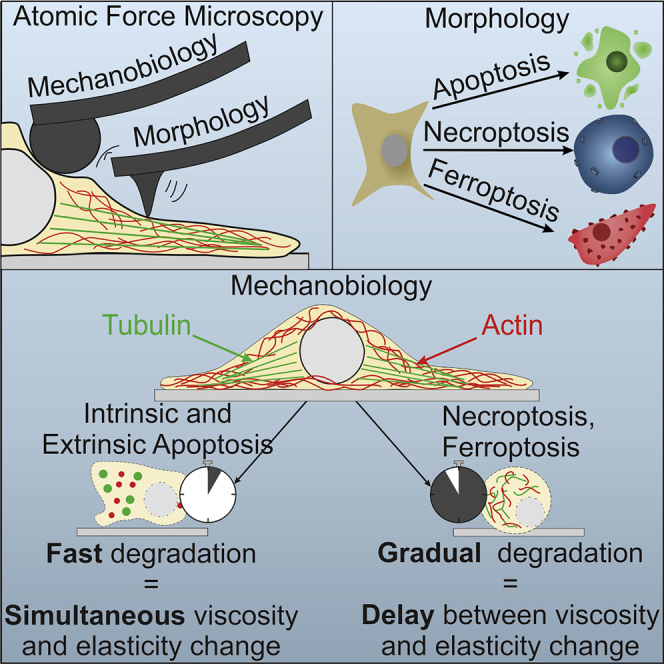
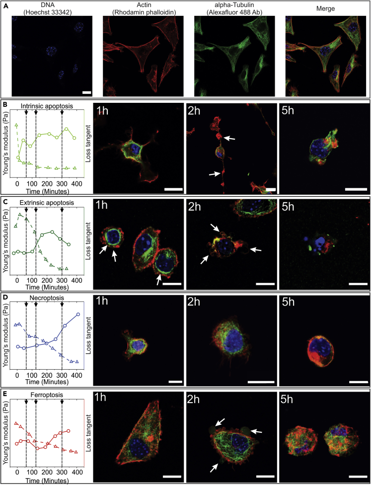
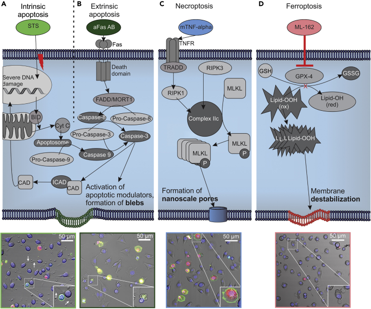
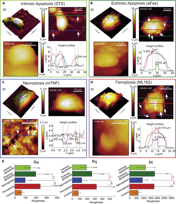
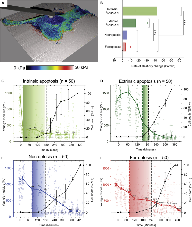
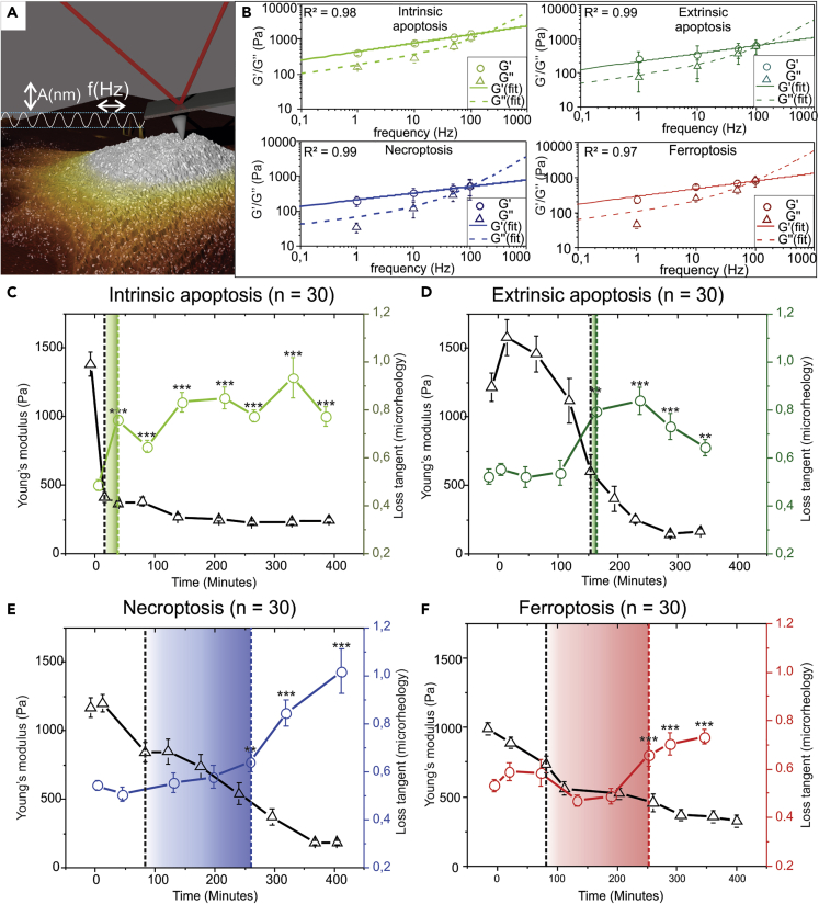
Regulated cell death (RCD) has a fundamental role in development, pathology, and tissue homeostasis. In order to understand the RCD mechanisms, it is essential to follow these processes in real time. Here, atomic force microscopy (AFM) is applied to morphologically and mechanically characterize four RCD modalities (intrinsic and extrinsic apoptosis, necroptosis, and ferroptosis) in murine tumor cell lines. The nano-topographical analysis revealed a distinct surface morphology in case of necroptosis, ∼ 200 nm membrane disruptions are observed. Using mechanical measurements, it is possible to detect the early onset of RCD. Combined elasticity and microrheology analysis allowed for a clear distinction between apoptotic and regulated necrotic cell death. Finally, immunofluorescence analysis of the cytoskeleton structure during the RCD processes confirm the measured mechanical changes. The results of this study not only demonstrate the possibility of early real-time cell death detection but also reveal important differences in the cytoskeletal dynamics between multiple RCD modalities.
程序性细胞死亡(RCD)在发育、病理学和组织稳态中起着至关重要的作用。为了理解RCD机制,实时跟踪这些过程至关重要。在此,原子力显微镜(AFM)被用于对小鼠肿瘤细胞系中的四种RCD模式(内源性和外源性凋亡、坏死性凋亡和铁死亡)进行形态学和力学表征。纳米拓扑分析显示,在坏死性凋亡的情况下,表面形态明显不同,观察到约200纳米的膜破坏。通过力学测量,可以检测到RCD的早期发作。弹性和微观流变学分析相结合,能够清楚地区分凋亡性和程序性坏死性细胞死亡。最后,对RCD过程中细胞骨架结构的免疫荧光分析证实了所测量的力学变化。本研究结果不仅证明了早期实时检测细胞死亡的可能性,还揭示了多种RCD模式之间细胞骨架动力学的重要差异。