Nuffield Department of Clinical Neurosciences, John Radcliffe Hospital, University of Oxford, Oxford, UK.
Nuffield Department of Medicine, Centre for Medicines Discovery, Target Discovery Institute, University of Oxford, Headington, UK.
Brain Pathol. 2021 Jul;31(4):e12923. doi: 10.1111/bpa.12923. Epub 2021 Jan 29.
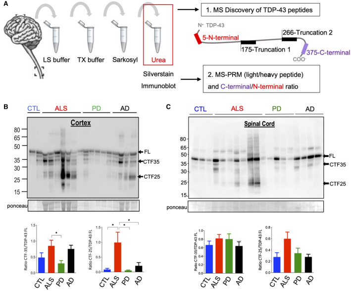
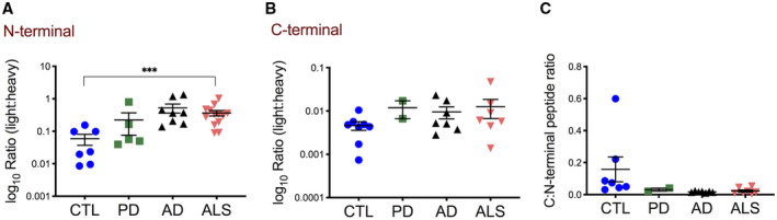
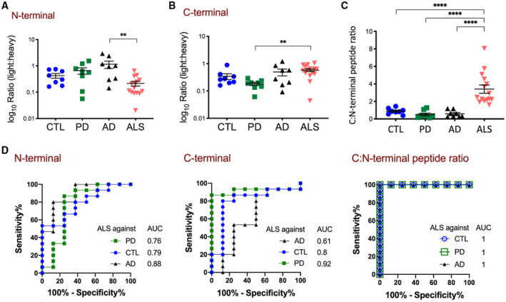
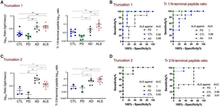
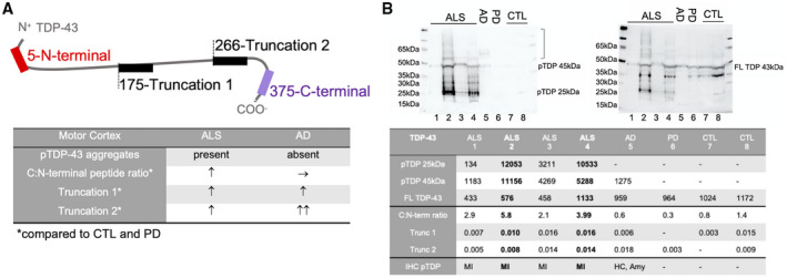
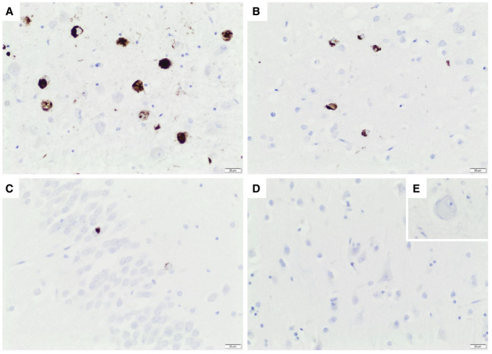
The pathological hallmark of amyotrophic lateral sclerosis (ALS) is the presence of cytoplasmic inclusions, containing C-terminal fragments of the protein TDP-43. Here, we tested the hypothesis that highly sensitive mass spectrometry with parallel reaction monitoring (MS-PRM) can generate a high-resolution map of pathological TDP-43 peptide ratios to form the basis for quantitation of abnormal C-terminal TDP-43 fragment enrichment. Human cortex and spinal cord, microscopically staged for the presence of p-TDP-43, p-tau, alpha-synuclein, and beta-amyloid pathology, were biochemically fractionated and analyzed by immunoblot and MS for the detection of full-length and truncated (disease-specific) TDP-43 peptides. This informed the synthesis of heavy isotope-labeled peptides for absolute quantification of TDP-43 by MS-PRM across 16 ALS, 8 Parkinson's, 8 Alzheimer's disease, and 8 aged control cases. We confirmed by immunoblot the previously described enrichment of pathological C-terminal fragments in ALS-TDP urea fractions. Subsequent MS analysis resolved specific TDP-43 N- and C-terminal peptides, including a novel N-terminal truncation site-specific peptide. Absolute quantification of peptides by MS-PRM showed an increased C:N-terminal TDP-43 peptide ratio in ALS-TDP brain compared to normal and disease controls. A C:N-terminal ratio >1.5 discriminated ALS from controls with a sensitivity of 100% (CI 79.6-100) and specificity of 100% (CI 68-100), and from Parkinson's and Alzheimer's disease with a sensitivity of 93% (CI 70-100) and specificity of 100% (CI 68-100). N-terminal truncation site-specific peptides were increased in ALS in line with C-terminal fragment enrichment, but were also found in a proportion of Alzheimer cases with normal C:N-terminal ratio but coexistent limbic TDP-43 neuropathological changes. In conclusion this is a novel, sensitive, and specific method to quantify the enrichment of pathological TDP-43 fragments in human brain, which could form the basis for an antibody-free assay. Our methodology has the potential to help clarify if specific pathological TDP-43 peptide signatures are associated with primary or secondary TDP-43 proteinopathies.
肌萎缩侧索硬化症(ALS)的病理学标志是细胞质包含物的存在,其中包含 TDP-43 蛋白的 C 末端片段。在这里,我们测试了以下假设:高灵敏度的质谱联用平行反应监测(MS-PRM)可以生成病理性 TDP-43 肽比的高分辨率图谱,为定量异常 C 末端 TDP-43 片段富集奠定基础。对显微镜下存在 p-TDP-43、p-tau、α-突触核蛋白和β-淀粉样蛋白病变的人脑皮质和脊髓进行生化分级,并通过免疫印迹和 MS 进行全长和截断(疾病特异性)TDP-43 肽的检测。这为通过 MS-PRM 对 16 例 ALS、8 例帕金森病、8 例阿尔茨海默病和 8 例老年对照组进行 TDP-43 的绝对定量合成重同位素标记肽提供了信息。我们通过免疫印迹证实了先前描述的 ALS-TDP 脲素级分中病理性 C 末端片段的富集。随后的 MS 分析解析了特定的 TDP-43 N-和 C-末端肽,包括一种新的 N-末端截断位点特异性肽。通过 MS-PRM 对肽的绝对定量显示,与正常和疾病对照相比,ALS-TDP 脑的 C:N-末端 TDP-43 肽比增加。C:N-末端比>1.5 可将 ALS 与对照组区分开来,其灵敏度为 100%(CI 79.6-100),特异性为 100%(CI 68-100),与帕金森病和阿尔茨海默病的灵敏度为 93%(CI 70-100),特异性为 100%(CI 68-100)。与 C 末端片段富集一致,ALS 中 N 末端截断位点特异性肽增加,但在一部分具有正常 C:N-末端比但共存边缘 TDP-43 神经病理学改变的阿尔茨海默病病例中也发现了这些肽。总之,这是一种新颖、敏感和特异的方法,可以定量人类大脑中病理性 TDP-43 片段的富集,这可能为无抗体测定奠定基础。我们的方法有可能帮助阐明特定的病理性 TDP-43 肽特征是否与原发性或继发性 TDP-43 蛋白病有关。