• 文献检索
  • 文档翻译
  • 深度研究
  • 学术资讯
  • Suppr Zotero 插件Zotero 插件
  • 邀请有礼
  • 套餐&价格
  • 历史记录
应用&插件
Suppr Zotero 插件Zotero 插件浏览器插件Mac 客户端Windows 客户端微信小程序
定价
高级版会员购买积分包购买API积分包
服务
文献检索文档翻译深度研究API 文档MCP 服务
关于我们
关于 Suppr公司介绍联系我们用户协议隐私条款
关注我们

Suppr 超能文献

核心技术专利:CN118964589B侵权必究
粤ICP备2023148730 号-1Suppr @ 2026

文献检索

告别复杂PubMed语法,用中文像聊天一样搜索,搜遍4000万医学文献。AI智能推荐,让科研检索更轻松。

立即免费搜索

文件翻译

保留排版,准确专业,支持PDF/Word/PPT等文件格式,支持 12+语言互译。

免费翻译文档

深度研究

AI帮你快速写综述,25分钟生成高质量综述,智能提取关键信息,辅助科研写作。

立即免费体验

光学显微镜和新型抗酸染色检测病变内结核分枝杆菌感染的三维原位形态计量学

Three-dimensional in situ morphometrics of Mycobacterium tuberculosis infection within lesions by optical mesoscopy and novel acid-fast staining.

机构信息

Biological Imaging Group, Analytical and Biological Sciences Division, National Institute for Biological Standards and Control (NIBSC), South Mimms, Hertfordshire, EN6 3QG, UK.

Department of Physics, SUPA, University of Strathclyde, 107 Rottenrow, Glasgow, G4 0NG, UK.

出版信息

Sci Rep. 2020 Dec 11;10(1):21774. doi: 10.1038/s41598-020-78640-4.

DOI:10.1038/s41598-020-78640-4
PMID:33311596
原文链接:https://pmc.ncbi.nlm.nih.gov/articles/PMC7733456/
Abstract

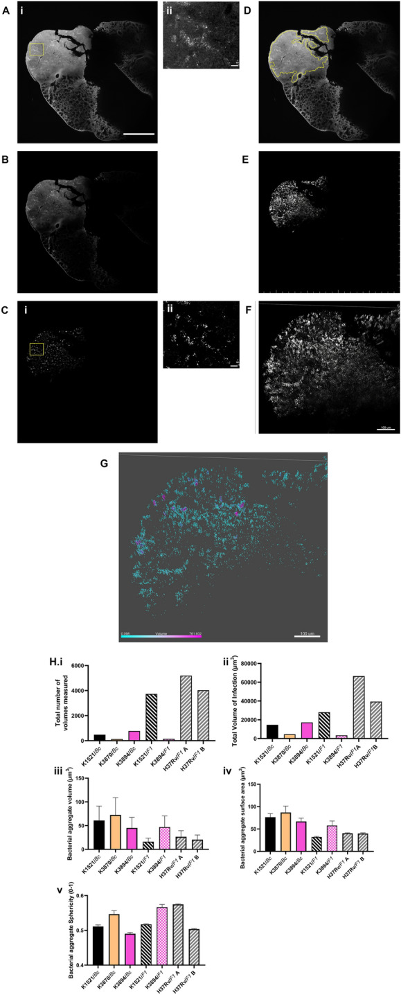
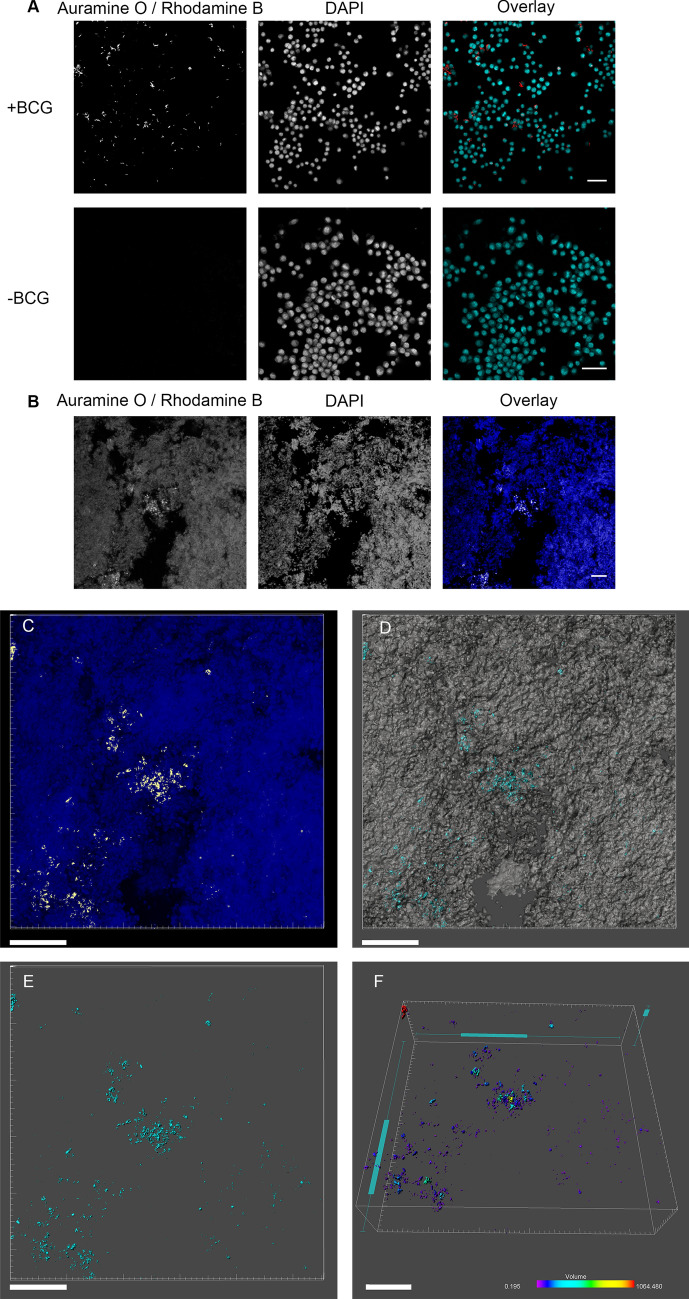
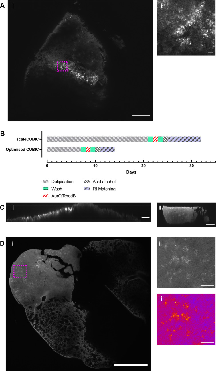
Tuberculosis (TB) preclinical testing relies on in vivo models including the mouse aerosol challenge model. The only method of determining colony morphometrics of TB infection in a tissue in situ is two-dimensional (2D) histopathology. 2D measurements consider heterogeneity within a single observable section but not above and below, which could contain critical information. Here we describe a novel approach, using optical clearing and a novel staining procedure with confocal microscopy and mesoscopy, for three-dimensional (3D) measurement of TB infection within lesions at sub-cellular resolution over a large field of view. We show TB morphometrics can be determined within lesion pathology, and differences in infection with different strains of Mycobacterium tuberculosis. Mesoscopy combined with the novel CUBIC Acid-Fast (CAF) staining procedure enables a quantitative approach to measure TB infection and allows 3D analysis of infection, providing a framework which could be used in the analysis of TB infection in situ.

摘要

结核病(TB)临床前检测依赖于体内模型,包括小鼠雾化挑战模型。确定组织内 TB 感染的菌落形态计量的唯一方法是二维(2D)组织病理学。2D 测量考虑了单个可观察切片内的异质性,但不考虑上下部分,因为上下部分可能包含关键信息。在这里,我们描述了一种新方法,使用光学透明化和共聚焦显微镜和体视学的新型染色程序,以亚细胞分辨率在大视场范围内对病变内的 TB 感染进行三维(3D)测量。我们表明可以在病变病理学中确定 TB 形态计量,并确定不同结核分枝杆菌菌株感染的差异。体视学结合新型立方酸快速(CAF)染色程序可以实现定量测量 TB 感染,并允许对感染进行 3D 分析,为原位 TB 感染分析提供了一个框架。

https://cdn.ncbi.nlm.nih.gov/pmc/blobs/0fc5/7733456/4b506d33d57e/41598_2020_78640_Fig4_HTML.jpg
https://cdn.ncbi.nlm.nih.gov/pmc/blobs/0fc5/7733456/6d6295f228cf/41598_2020_78640_Fig1_HTML.jpg
https://cdn.ncbi.nlm.nih.gov/pmc/blobs/0fc5/7733456/0f02d194a2f3/41598_2020_78640_Fig2_HTML.jpg
https://cdn.ncbi.nlm.nih.gov/pmc/blobs/0fc5/7733456/6b0e11dc3dea/41598_2020_78640_Fig3_HTML.jpg
https://cdn.ncbi.nlm.nih.gov/pmc/blobs/0fc5/7733456/4b506d33d57e/41598_2020_78640_Fig4_HTML.jpg

相似文献

1
Three-dimensional in situ morphometrics of Mycobacterium tuberculosis infection within lesions by optical mesoscopy and novel acid-fast staining.光学显微镜和新型抗酸染色检测病变内结核分枝杆菌感染的三维原位形态计量学
Sci Rep. 2020 Dec 11;10(1):21774. doi: 10.1038/s41598-020-78640-4.
2
Presence of multiple lesion types with vastly different microenvironments in C3HeB/FeJ mice following aerosol infection with Mycobacterium tuberculosis.用结核分枝杆菌气溶胶感染C3HeB/FeJ小鼠后,出现具有截然不同微环境的多种病变类型。
Dis Model Mech. 2015 Jun;8(6):591-602. doi: 10.1242/dmm.019570. Epub 2015 Mar 30.
3
Modified immunohistological staining allows detection of Ziehl-Neelsen-negative Mycobacterium tuberculosis organisms and their precise localization in human tissue.改良免疫组织化学染色可检测萋-尼氏染色阴性的结核分枝杆菌及其在人体组织中的精确定位。
J Pathol. 2005 Apr;205(5):633-40. doi: 10.1002/path.1728.
4
Mouse models of human TB pathology: roles in the analysis of necrosis and the development of host-directed therapies.人类结核病病理学的小鼠模型:在坏死分析和宿主导向疗法开发中的作用。
Semin Immunopathol. 2016 Mar;38(2):221-37. doi: 10.1007/s00281-015-0538-9. Epub 2015 Nov 5.
5
A comparative study on the different staining methods and number of specimens for the detection of acid fast bacilli.不同染色方法及标本数量对结核杆菌检测的比较研究
Mem Inst Oswaldo Cruz. 2000 Nov-Dec;95(6):855-8. doi: 10.1590/s0074-02762000000600019.
6
Nucleic Acid Amplification Testing and Sequencing Combined with Acid-Fast Staining in Needle Biopsy Lung Tissues for the Diagnosis of Smear-Negative Pulmonary Tuberculosis.核酸扩增检测与测序联合抗酸染色在经皮肺穿刺活检组织中诊断涂片阴性肺结核的应用
PLoS One. 2016 Dec 2;11(12):e0167342. doi: 10.1371/journal.pone.0167342. eCollection 2016.
7
[Recent progress in mycobacteriology].[分枝杆菌学的最新进展]
Kekkaku. 2007 Oct;82(10):783-99.
8
Immunohistochemical localization of mycobacterium tuberculosis complex antigen with antibody to 38 kDa antigen versus Ziehl Neelsen staining in tissue granulomas of extrapulmonary tuberculosis.在肺外结核组织肉芽肿中,用抗38 kDa抗原抗体对结核分枝杆菌复合抗原进行免疫组织化学定位与齐-尼氏染色的比较
Indian J Tuberc. 2007 Jan;54(1):24-9.
9
Correlative light electron ion microscopy reveals in vivo localisation of bedaquiline in Mycobacterium tuberculosis-infected lungs.相关光电子离子显微镜揭示了体内利奈唑胺在感染结核分枝杆菌的肺部中的定位。
PLoS Biol. 2020 Dec 31;18(12):e3000879. doi: 10.1371/journal.pbio.3000879. eCollection 2020 Dec.
10
Micro-PET imaging of [18F]fluoroacetate combined with [18F]FDG to differentiate chronic Mycobacterium tuberculosis infection from an acute bacterial infection in a mouse model: a preliminary study.[18F]氟乙酸盐联合[18F]氟代脱氧葡萄糖的微型正电子发射断层扫描成像在小鼠模型中鉴别慢性结核分枝杆菌感染与急性细菌感染的初步研究
Nucl Med Commun. 2019 Jun;40(6):639-644. doi: 10.1097/MNM.0000000000001017.

引用本文的文献

1
Correlative 3D imaging method for analysing lesion architecture in susceptible mice infected with Mycobacterium tuberculosis.用于分析感染结核分枝杆菌的易感小鼠病变结构的相关三维成像方法。
Dis Model Mech. 2025 Sep 1;18(9). doi: 10.1242/dmm.052185. Epub 2025 Mar 26.
2
Current understanding of the human microbiome in glioma.当前对胶质瘤中人类微生物组的认识。
Front Oncol. 2022 Aug 8;12:781741. doi: 10.3389/fonc.2022.781741. eCollection 2022.
3
Application of Light-Sheet Mesoscopy to Image Host-Pathogen Interactions in Intact Organs.

本文引用的文献

1
Application of the Mesolens for subcellular resolution imaging of intact larval and whole adult Drosophila.Mesolens 用于完整幼虫和成年果蝇的亚细胞分辨率成像的应用。
J Microsc. 2018 May;270(2):252-258. doi: 10.1111/jmi.12693. Epub 2018 Mar 23.
2
A three-dimensional single-cell-resolution whole-brain atlas using CUBIC-X expansion microscopy and tissue clearing.采用 CUBIC-X 扩展显微镜和组织透明化技术的三维单细胞分辨率全脑图谱
Nat Neurosci. 2018 Apr;21(4):625-637. doi: 10.1038/s41593-018-0109-1. Epub 2018 Mar 5.
3
Quantifying intracellular Mycobacterium tuberculosis: An essential issue for in vitro assays.
应用光片介观成像技术观察完整器官内的宿主-病原体相互作用。
Front Cell Infect Microbiol. 2022 Jun 14;12:903957. doi: 10.3389/fcimb.2022.903957. eCollection 2022.
4
Challenges and advances in optical 3D mesoscale imaging.光学 3D 介观成像的挑战与进展。
J Microsc. 2022 Jun;286(3):201-219. doi: 10.1111/jmi.13109. Epub 2022 May 5.
定量胞内结核分枝杆菌:体外检测的重要问题。
Microbiologyopen. 2018 Apr;7(2):e00588. doi: 10.1002/mbo3.588. Epub 2018 Feb 27.
4
CUBIC pathology: three-dimensional imaging for pathological diagnosis.立方病理:用于病理诊断的三维成像。
Sci Rep. 2017 Aug 24;7(1):9269. doi: 10.1038/s41598-017-09117-0.
5
Whole-Body Profiling of Cancer Metastasis with Single-Cell Resolution.以单细胞分辨率进行癌症转移的全身分析
Cell Rep. 2017 Jul 5;20(1):236-250. doi: 10.1016/j.celrep.2017.06.010.
6
Hypervirulent Mycobacterium tuberculosis strain triggers necrotic lung pathology associated with enhanced recruitment of neutrophils in resistant C57BL/6 mice.高毒力结核分枝杆菌菌株在抗性C57BL/6小鼠中引发与中性粒细胞募集增加相关的坏死性肺部病理变化。
PLoS One. 2017 Mar 17;12(3):e0173715. doi: 10.1371/journal.pone.0173715. eCollection 2017.
7
Genomic diversity in autopsy samples reveals within-host dissemination of HIV-associated Mycobacterium tuberculosis.尸检样本中的基因组多样性揭示了HIV相关结核分枝杆菌在宿主体内的播散。
Nat Med. 2016 Dec;22(12):1470-1474. doi: 10.1038/nm.4205. Epub 2016 Oct 31.
8
A novel optical microscope for imaging large embryos and tissue volumes with sub-cellular resolution throughout.一种新型光学显微镜,可对大型胚胎和组织进行整体亚细胞分辨率成像。
Elife. 2016 Sep 23;5:e18659. doi: 10.7554/eLife.18659.
9
Chemical Principles in Tissue Clearing and Staining Protocols for Whole-Body Cell Profiling.化学原理在组织通透和染色技术中的应用,用于整体细胞分析。
Annu Rev Cell Dev Biol. 2016 Oct 6;32:713-741. doi: 10.1146/annurev-cellbio-111315-125001. Epub 2016 Jun 8.
10
The Importance of First Impressions: Early Events in Mycobacterium tuberculosis Infection Influence Outcome.第一印象的重要性:结核分枝杆菌感染早期事件影响结局
mBio. 2016 Apr 5;7(2):e00342-16. doi: 10.1128/mBio.00342-16.