Division of Gastroenterology and Hepatology, Department of Internal Medicine, National Taiwan University Hospital, Taipei, Taiwan.
Division of Gastroenterology and Hepatology, Department of Internal Medicine, National Taiwan University Hospital Bei-Hu Branch, Taipei, Taiwan.
Clin Mol Hepatol. 2021 Apr;27(2):305-312. doi: 10.3350/cmh.2020.0301. Epub 2020 Dec 3.
BACKGROUND/AIMS: The core needle biopsy (CNB), fine needle aspiration cytology (FNAC) and touch imprint cytology (TIC) are commonly used tools for the diagnosis of hepatic malignancies. However, little is known about the benefits and criteria for selecting appropriate technique among them in clinical practice. We aimed to compare the sensitivity of ultrasound-guided CNB, FNAC, TIC as well as combinations for the diagnosis of hepatic malignancies, and to determine the factors associated with better sensitivity in each technique.
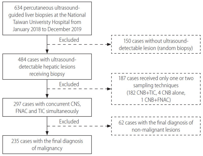
From January 2018 to December 2019, a total of 634 consecutive patients who received ultrasound-guided liver biopsies at the National Taiwan University Hospital was collected, of whom 235 with confirmed malignant hepatic lesions receiving CNB, FNAC and TIC simultaneously were enrolled for analysis. The clinical and procedural data were compared.
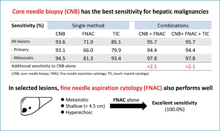
The sensitivity of CNB, FNAC and TIC for the diagnosis of malignant hepatic lesions were 93.6%, 71.9%, and 85.1%, respectively. Add-on use of FNAC or TIC to CNB provided additional sensitivity of 2.1% and 0.4%, respectively. FNAC exhibited a significantly higher diagnostic rate in the metastatic cancers (P=0.011), hyperechoic lesions on ultrasound (P=0.028), and those with depth less than 4.5 cm from the site of needle insertion (P=0.036).
The sensitivity of CNB is superior to that of FNAC and TIC for the diagnosis of hepatic malignancies. Nevertheless, for shallow (depth <4.5 cm) and hyperechoic lesions not typical for primary liver cancers, FNAC alone provides excellent sensitivity.
背景/目的: 核心针活检 (CNB)、细针抽吸细胞学 (FNAC) 和触诊印片细胞学 (TIC) 是诊断肝恶性肿瘤的常用工具。然而,在临床实践中,对于选择其中合适技术的益处和标准知之甚少。我们旨在比较超声引导下 CNB、FNAC、TIC 以及它们的组合在诊断肝恶性肿瘤方面的敏感性,并确定每种技术中与更好敏感性相关的因素。
2018 年 1 月至 2019 年 12 月,共收集了在台湾大学医院接受超声引导下肝活检的 634 例连续患者,其中 235 例经证实患有恶性肝病变的患者同时接受了 CNB、FNAC 和 TIC 检查,对其进行了分析。比较了临床和程序数据。
CNB、FNAC 和 TIC 诊断恶性肝病变的敏感性分别为 93.6%、71.9%和 85.1%。在 CNB 上加用 FNAC 或 TIC 可分别提供 2.1%和 0.4%的额外敏感性。FNAC 在转移性癌症 (P=0.011)、超声上高回声病变 (P=0.028) 和距离针插入部位小于 4.5 厘米的病变中具有显著更高的诊断率 (P=0.036)。
CNB 诊断肝恶性肿瘤的敏感性优于 FNAC 和 TIC。然而,对于不典型的原发性肝癌的浅层 (深度 <4.5 厘米) 和高回声病变,单独使用 FNAC 可提供出色的敏感性。