Barnes Steven, Grove James C R, McHugh Cyrus F, Hirano Arlene A, Brecha Nicholas C
Doheny Eye Institute, Los Angeles, CA, United States.
Department of Ophthalmology, David Geffen School of Medicine, University of California, Los Angeles, Los Angeles, CA, United States.
Front Cell Neurosci. 2020 Nov 4;14:595064. doi: 10.3389/fncel.2020.595064. eCollection 2020.
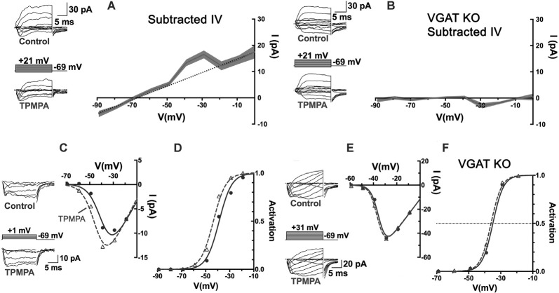
How neurons in the eye feed signals back to photoreceptors to optimize sensitivity to patterns of light appears to be mediated by one or more unconventional mechanisms. Via these mechanisms, horizontal cells control photoreceptor synaptic gain and enhance key aspects of temporal and spatial center-surround receptive field antagonism. After the transduction of light energy into an electrical signal in photoreceptors, the next key task in visual processing is the transmission of an optimized signal to the follower neurons in the retina. For this to happen, the release of the excitatory neurotransmitter glutamate from photoreceptors is carefully regulated via horizontal cell feedback, which acts as a thermostat to keep the synaptic transmission in an optimal range during changes to light patterns and intensities. Novel findings of a recently described model that casts a classical neurotransmitter system together with ion transport mechanisms to adjust the alkaline milieu outside the synapse are reviewed. This novel inter-neuronal messaging system carries feedback signals using two separate, but interwoven regulated systems. The complex interplay between these two signaling modalities, creating synaptic modulation-at-a-distance, has obscured it's being defined. The foundations of our understanding of the feedback mechanism from horizontal cells to photoreceptors have been long established: Horizontal cells have broad receptive fields, suitable for providing surround inhibition, their membrane potential, a function of stimulus intensity and size, regulates inhibition of photoreceptor voltage-gated Ca channels, and strong artificial pH buffering eliminates this action. This review compares and contrasts models of how these foundations are linked, focusing on a recent report in mammals that shows tonic horizontal cell release of GABA activating Cl and HCO permeable GABA autoreceptors. The membrane potential of horizontal cells provides the driving force for GABAR-mediated HCO efflux, alkalinizing the cleft when horizontal cells are hyperpolarized by light or adding to their depolarization in darkness and contributing to cleft acidification NHE-mediated H efflux. This model challenges interpretations of earlier studies that were considered to rule out a role for GABA in feedback to cones.
眼睛中的神经元如何将信号反馈给光感受器以优化对光模式的敏感度,这似乎是由一种或多种非常规机制介导的。通过这些机制,水平细胞控制光感受器的突触增益,并增强时间和空间中心-外周感受野拮抗作用的关键方面。在光感受器将光能转化为电信号之后,视觉处理中的下一个关键任务是将优化后的信号传递给视网膜中的后续神经元。为此,光感受器释放兴奋性神经递质谷氨酸的过程会通过水平细胞反馈进行精细调节,水平细胞反馈就像一个恒温器,在光模式和强度发生变化时,将突触传递保持在最佳范围内。本文综述了最近描述的一个模型的新发现,该模型将经典神经递质系统与离子转运机制结合起来,以调节突触外的碱性环境。这个新的神经元间信息传递系统使用两个独立但相互交织的调节系统来携带反馈信号。这两种信号传导方式之间的复杂相互作用产生了远距离的突触调制,这使得对它的定义变得模糊不清。我们对从水平细胞到光感受器的反馈机制的理解基础早已确立:水平细胞具有广泛的感受野,适合提供外周抑制,其膜电位是刺激强度和大小的函数,调节对光感受器电压门控钙通道的抑制,并且强大的人工pH缓冲会消除这种作用。本综述比较并对比了这些基础如何相互关联的模型,重点关注哺乳动物中最近的一份报告,该报告显示水平细胞持续释放GABA,激活Cl和HCO可渗透的GABA自身受体。水平细胞的膜电位为GABAR介导的HCO外流提供驱动力,当水平细胞因光而超极化时使突触间隙碱化,或在黑暗中增加其去极化,并促进由NHE介导的H外流导致的突触间隙酸化。该模型挑战了早期研究的解释,这些研究被认为排除了GABA在向视锥细胞反馈中的作用。