• 文献检索
  • 文档翻译
  • 深度研究
  • 学术资讯
  • Suppr Zotero 插件Zotero 插件
  • 邀请有礼
  • 套餐&价格
  • 历史记录
应用&插件
Suppr Zotero 插件Zotero 插件浏览器插件Mac 客户端Windows 客户端微信小程序
定价
高级版会员购买积分包购买API积分包
服务
文献检索文档翻译深度研究API 文档MCP 服务
关于我们
关于 Suppr公司介绍联系我们用户协议隐私条款
关注我们

Suppr 超能文献

核心技术专利:CN118964589B侵权必究
粤ICP备2023148730 号-1Suppr @ 2026

文献检索

告别复杂PubMed语法,用中文像聊天一样搜索,搜遍4000万医学文献。AI智能推荐,让科研检索更轻松。

立即免费搜索

文件翻译

保留排版,准确专业,支持PDF/Word/PPT等文件格式,支持 12+语言互译。

免费翻译文档

深度研究

AI帮你快速写综述,25分钟生成高质量综述,智能提取关键信息,辅助科研写作。

立即免费体验

冠心病患者高切变应力与 OCT 验证的薄帽纤维粥样瘤之间的关系。

Relationship between high shear stress and OCT-verified thin-cap fibroatheroma in patients with coronary artery disease.

机构信息

Division of Cardiology, Mount Sinai Hospital and Icahn School of Medicine at Mount Sinai, New York, NY, United States of America.

Division of Cardiology, Department of Medicine, Emory University School of Medicine, Atlanta, GA, United States of America.

出版信息

PLoS One. 2020 Dec 17;15(12):e0244015. doi: 10.1371/journal.pone.0244015. eCollection 2020.

DOI:10.1371/journal.pone.0244015
PMID:33332434
原文链接:https://pmc.ncbi.nlm.nih.gov/articles/PMC7746187/
Abstract

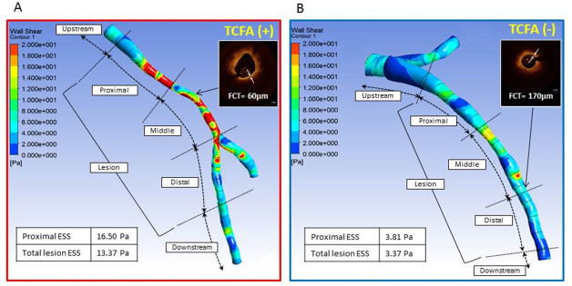
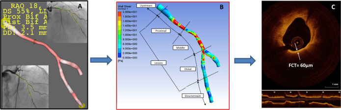
High-risk coronary plaques have been considered predictive of adverse cardiac events. Both wall shear stress (WSS) in patients with hemodynamically significant lesions and optical coherence tomography (OCT) -verified thin-cap fibroatheroma (TCFA) are associated with plaque rupture, the most common underlying mechanism of acute coronary syndrome. The aim of the study was to test the hypothesis that invasive coronary angiography-based high WSS is associated with the presence of TCFA detected by OCT in obstructive lesions. From a prospective study of patients who underwent OCT examination for angiographically obstructive lesions (Yellow II), we selected patients who had two angiographic projections to create a 3-dimensional reconstruction model to allow assessment of WSS. The patients were divided into 2 groups according to the presence and absence of TCFA. Mean WSS was assessed in the whole lesion and in the proximal, middle and distal segments. Of 70 patients, TCFA was observed in 13 (19%) patients. WSS in the proximal segment (WSSproximal) (10.20 [5.01, 16.93Pa]) and the whole lesion (WSSlesion) (12.37 [6.36, 14.55Pa]) were significantly higher in lesions with TCFA compared to WSSproximal (5.84 [3.74, 8.29Pa], p = 0.02) and WSSlesion (6.95 [4.41, 11.60], p = 0.04) in lesions without TCFA. After multivariate analysis, WSSproximal was independently associated with the presence of TCFA (Odds ratio 1.105; 95%CI 1.007-1.213, p = 0.04). The optimal cutoff value of WSSproximal to predict TCFA was 6.79 Pa (AUC: 0.71; sensitivity: 0.77; specificity: 0.63 p = 0.02). Our results demonstrate that high WSS in the proximal segments of obstructive lesions is an independent predictor of OCT-verified TCFA.

摘要

高危冠状动脉斑块一直被认为是预测不良心脏事件的指标。血流动力学意义上的病变中的壁面切应力(WSS)和光学相干断层扫描(OCT)证实的薄帽纤维粥样斑块(TCFA)都与斑块破裂有关,这是急性冠脉综合征最常见的潜在机制。本研究的目的是检验假设,即基于侵入性冠状动脉造影的高壁面切应力(WSS)与 OCT 检测到的阻塞性病变中的 TCFA 存在相关。我们从接受 OCT 检查的血管造影阻塞性病变(Yellow II)的前瞻性研究中选择患者,这些患者有两个血管造影投影来创建 3 维重建模型,以评估 WSS。根据是否存在 TCFA,将患者分为两组。评估整个病变和近端、中段和远端节段的平均 WSS。在 70 例患者中,13 例(19%)患者观察到 TCFA。与无 TCFA 的病变相比,TCFA 病变的近端节段(WSSproximal)(10.20[5.01,16.93Pa])和整个病变(WSSlesion)(12.37[6.36,14.55Pa])的 WSS 更高(WSSproximal 为 5.84[3.74,8.29Pa],p=0.02;WSSlesion 为 6.95[4.41,11.60],p=0.04)。多变量分析后,WSSproximal 与 TCFA 的存在独立相关(比值比 1.105;95%CI 1.007-1.213,p=0.04)。预测 TCFA 的 WSSproximal 最佳截断值为 6.79Pa(AUC:0.71;敏感性:0.77;特异性:0.63,p=0.02)。我们的结果表明,阻塞性病变近端节段的高 WSS 是 OCT 证实的 TCFA 的独立预测因子。

https://cdn.ncbi.nlm.nih.gov/pmc/blobs/9950/7746187/f283a4f9e987/pone.0244015.g002.jpg
https://cdn.ncbi.nlm.nih.gov/pmc/blobs/9950/7746187/fabd67eb5e45/pone.0244015.g001.jpg
https://cdn.ncbi.nlm.nih.gov/pmc/blobs/9950/7746187/f283a4f9e987/pone.0244015.g002.jpg
https://cdn.ncbi.nlm.nih.gov/pmc/blobs/9950/7746187/fabd67eb5e45/pone.0244015.g001.jpg
https://cdn.ncbi.nlm.nih.gov/pmc/blobs/9950/7746187/f283a4f9e987/pone.0244015.g002.jpg

相似文献

1
Relationship between high shear stress and OCT-verified thin-cap fibroatheroma in patients with coronary artery disease.冠心病患者高切变应力与 OCT 验证的薄帽纤维粥样瘤之间的关系。
PLoS One. 2020 Dec 17;15(12):e0244015. doi: 10.1371/journal.pone.0244015. eCollection 2020.
2
Coronary CT angiography characteristics of OCT-defined thin-cap fibroatheroma: a section-to-section comparison study.OCT 定义的薄帽纤维粥样瘤的冠状动脉 CT 血管造影特征:一项节段到节段比较研究。
Eur Radiol. 2018 Feb;28(2):833-843. doi: 10.1007/s00330-017-4992-8. Epub 2017 Aug 23.
3
Atherosclerotic plaque characterization by CT angiography for identification of high-risk coronary artery lesions: a comparison to optical coherence tomography.通过CT血管造影对动脉粥样硬化斑块进行特征分析以识别高危冠状动脉病变:与光学相干断层扫描的比较
Eur Heart J Cardiovasc Imaging. 2015 Apr;16(4):373-9. doi: 10.1093/ehjci/jeu188. Epub 2014 Sep 21.
4
Thin-Cap Fibroatheroma Rather Than Any Lipid Plaques Increases the Risk of Cardiovascular Events in Diabetic Patients: Insights From the COMBINE OCT-FFR Trial.薄帽纤维粥样斑块而非任何脂质斑块增加糖尿病患者心血管事件的风险:来自 COMBINE OCT-FFR 试验的见解。
Circ Cardiovasc Interv. 2022 May;15(5):e011728. doi: 10.1161/CIRCINTERVENTIONS.121.011728. Epub 2022 Apr 29.
5
Plaque characteristics of thin-cap fibroatheroma evaluated by OCT and IVUS.OCT 和 IVUS 评价的薄帽纤维粥样斑块特征。
JACC Cardiovasc Imaging. 2011 Jun;4(6):638-46. doi: 10.1016/j.jcmg.2011.03.014.
6
Elevated levels of systemic pentraxin 3 are associated with thin-cap fibroatheroma in coronary culprit lesions: assessment by optical coherence tomography and intravascular ultrasound.循环系统 pentraxin 3 水平升高与冠状动脉罪犯病变中的薄帽纤维粥样瘤相关:光学相干断层成像和血管内超声评估。
JACC Cardiovasc Interv. 2013 Sep;6(9):945-54. doi: 10.1016/j.jcin.2013.04.024. Epub 2013 Aug 14.
7
Prevalence and predictors of culprit plaque rupture at OCT in patients with coronary artery disease: a meta-analysis.冠心病患者 OCT 检测中罪犯斑块破裂的患病率及预测因素:一项荟萃分析。
Eur Heart J Cardiovasc Imaging. 2016 Oct;17(10):1128-37. doi: 10.1093/ehjci/jev283. Epub 2015 Oct 27.
8
Feasibility of noninvasive assessment of thin-cap fibroatheroma by multidetector computed tomography.多排螺旋 CT 无创评估薄帽纤维粥样瘤的可行性。
JACC Cardiovasc Imaging. 2009 Dec;2(12):1412-9. doi: 10.1016/j.jcmg.2009.09.012.
9
Long-term outcomes of patients with normal fractional flow reserve and thin-cap fibroatheroma.正常分数血流储备患者和薄帽纤维粥样斑块患者的长期预后。
EuroIntervention. 2023 Feb 6;18(13):e1099-e1107. doi: 10.4244/EIJ-D-22-00306.
10
Computed Tomography Angiography Characteristics of Thin-Cap Fibroatheroma in Patients With Diabetes.糖尿病患者薄帽纤维粥样瘤的计算机断层血管造影特征。
J Am Heart Assoc. 2024 May 21;13(10):e033639. doi: 10.1161/JAHA.123.033639. Epub 2024 May 14.

引用本文的文献

1
The Role of Fluid Mechanics in Coronary Atherosclerotic Plaques: An Up-to-Date Review.流体力学在冠状动脉粥样硬化斑块中的作用:最新综述
Rev Cardiovasc Med. 2024 Jan 29;25(2):49. doi: 10.31083/j.rcm2502049. eCollection 2024 Feb.
2
Coronary Physiology-Based Approaches for Plaque Vulnerability: Implications for Risk Prediction and Treatment Strategies.基于冠状动脉生理学的斑块易损性评估方法:对风险预测和治疗策略的启示
Korean Circ J. 2023 Sep;53(9):581-593. doi: 10.4070/kcj.2023.0117.
3
Decay of Skin-Specific Gene Modules in Pangolins.穿山甲皮肤特异性基因模块的衰减。

本文引用的文献

1
Lipid-rich Plaques Detected by Near-infrared Spectroscopy Are More Frequently Exposed to High Shear Stress.近红外光谱检测到富含脂质的斑块更频繁地暴露于高剪切应力下。
J Cardiovasc Transl Res. 2021 Jun;14(3):416-425. doi: 10.1007/s12265-020-10072-x. Epub 2020 Oct 9.
2
Wall Shear Stress and Plaque Vulnerability: Computational Fluid Dynamics Analysis Derived From cCTA and OCT.壁面剪应力与斑块易损性:源自cCTA和OCT的计算流体动力学分析
JACC Cardiovasc Imaging. 2021 Jan;14(1):315-317. doi: 10.1016/j.jcmg.2020.07.034. Epub 2020 Sep 16.
3
Shear Stress Estimated by Quantitative Coronary Angiography Predicts Plaques Prone to Progress and Cause Events.
J Mol Evol. 2023 Aug;91(4):458-470. doi: 10.1007/s00239-023-10118-z. Epub 2023 May 30.
4
Atherosclerosis and endothelial mechanotransduction: current knowledge and models for future research.动脉粥样硬化与血管内皮细胞力学转导:当前知识与未来研究模型。
Am J Physiol Cell Physiol. 2023 Feb 1;324(2):C488-C504. doi: 10.1152/ajpcell.00449.2022. Epub 2022 Nov 28.
5
Investigation of Wall Shear Stress in Cardiovascular Research and in Clinical Practice-From Bench to Bedside.血管壁切应力在心血管研究和临床实践中的应用——从基础到临床。
Int J Mol Sci. 2021 May 26;22(11):5635. doi: 10.3390/ijms22115635.
定量冠状动脉造影估计的切应力可预测易进展和导致事件的斑块。
JACC Cardiovasc Imaging. 2020 Oct;13(10):2206-2219. doi: 10.1016/j.jcmg.2020.02.028. Epub 2020 May 13.
4
High shear stress on the coronary arterial wall is related to computed tomography-derived high-risk plaque: a three-dimensional computed tomography and color-coded tissue-characterizing intravascular ultrasonography study.冠状动脉壁上的高剪切应力与计算机断层扫描衍生的高危斑块相关:一项三维计算机断层扫描和彩色编码组织特征血管内超声检查研究。
Heart Vessels. 2019 Sep;34(9):1429-1439. doi: 10.1007/s00380-019-01389-y. Epub 2019 Apr 11.
5
Endothelial Shear Stress and Plaque Erosion: A Computational Fluid Dynamics and Optical Coherence Tomography Study.内皮剪切应力与斑块侵蚀:一项计算流体动力学和光学相干断层扫描研究
JACC Cardiovasc Imaging. 2019 Feb;12(2):374-375. doi: 10.1016/j.jcmg.2018.07.024. Epub 2018 Aug 28.
6
High Coronary Shear Stress in Patients With Coronary Artery Disease Predicts Myocardial Infarction.冠心病患者的高冠状动脉剪切应力可预测心肌梗死。
J Am Coll Cardiol. 2018 Oct 16;72(16):1926-1935. doi: 10.1016/j.jacc.2018.07.075.
7
Identification of High-Risk Plaques Destined to Cause Acute Coronary Syndrome Using Coronary Computed Tomographic Angiography and Computational Fluid Dynamics.采用冠状动脉计算机断层扫描血管造影和计算流体力学识别易致急性冠状动脉综合征的高危斑块。
JACC Cardiovasc Imaging. 2019 Jun;12(6):1032-1043. doi: 10.1016/j.jcmg.2018.01.023. Epub 2018 Mar 14.
8
Role of Low Endothelial Shear Stress and Plaque Characteristics in the Prediction of Nonculprit Major Adverse Cardiac Events: The PROSPECT Study.低内皮切应力和斑块特征在预测非罪犯主要不良心脏事件中的作用:PROSPECT 研究。
JACC Cardiovasc Imaging. 2018 Mar;11(3):462-471. doi: 10.1016/j.jcmg.2017.01.031. Epub 2017 Sep 18.
9
Near-infrared spectroscopy-derived lipid core burden index predicts adverse cardiovascular outcome in patients with coronary artery disease during long-term follow-up.近红外光谱衍生的脂质核心负荷指数可预测冠心病患者长期随访期间的不良心血管结局。
Eur Heart J. 2018 Jan 21;39(4):295-302. doi: 10.1093/eurheartj/ehx247.
10
Clinical Significance of Lipid-Rich Plaque Detected by Optical Coherence Tomography: A 4-Year Follow-Up Study.光学相干断层扫描检测富含脂质斑块的临床意义:一项为期 4 年的随访研究。
J Am Coll Cardiol. 2017 May 23;69(20):2502-2513. doi: 10.1016/j.jacc.2017.03.556.