Zheng Jun, Kuang Jing, Zhang Xianyu, Luo Daya, Liao Weijing
Department of Neurorehabilitation, Zhongnan Hospital of Wuhan University, Wuhan, Hubei, 430071, China.
Department of Plastic Surgery, Jinan Central Hospital Affiliated to Shandong University, Jinan, Shandong, 250013, China.
Transl Neurosci. 2020 May 18;11(1):105-115. doi: 10.1515/tnsci-2020-0105. eCollection 2020.
Spinal cord injury (SCI) leads to abnormal expression of miRs, leading to secondary responses such as oxidative stress, inflammation and apoptosis. In the present work, we screened the miRs involved and the associated pathway.
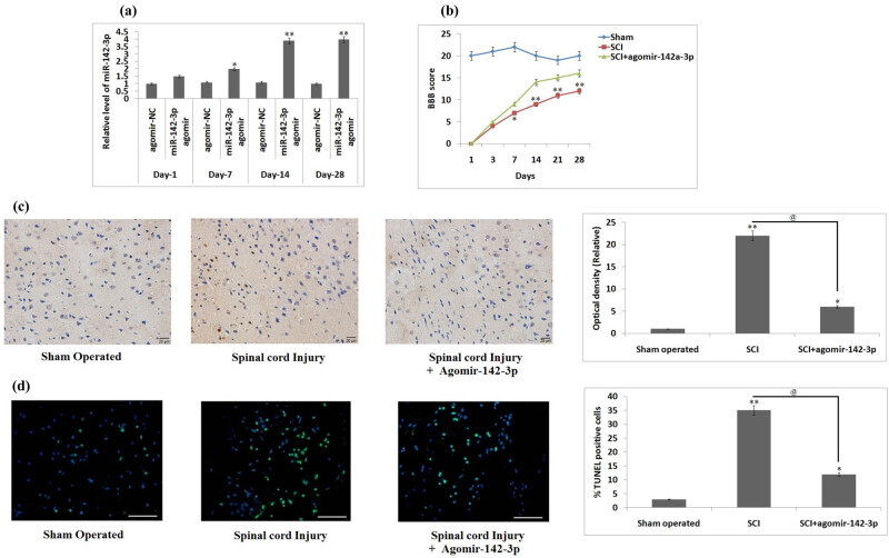
In a rat model of SCI, the microarray analysis for expression of miRs at various time points post-SCI was done. The locomotor analysis was done by Basso, Beattie and Bresnahan score, and Cresyl violet staining was done for lesion volume and TUNEL assay was done for apoptosis in neuronal cells. The expression of apoptotic proteins was done by the western blot study.
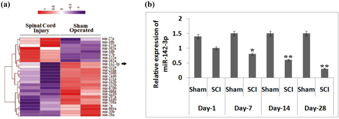
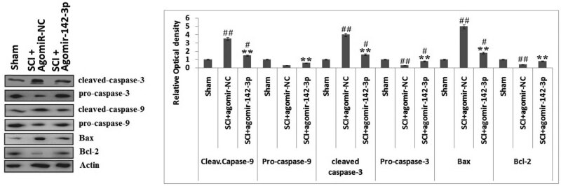
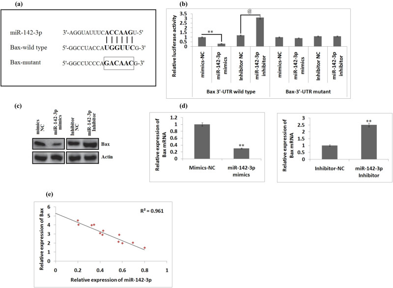
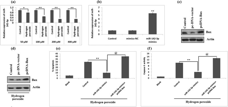
It was evidenced that the expression of the number of miRs was altered on the 14th day post-SCI, and miR-142-3p was found to be the most significantly suppressed miR. The results suggested that overexpression of miR-142-3p by its agomir-attenuated functional recovery decreased lesion size and apoptosis of neuronal cells in rats subjected to SCI. The luciferase assay indicated that miR-142-3p blocked the levels of Bax, which is a significant activator of the mitochondrial apoptotic pathway (MAP) via targeting the 3'UTR region of BV-2 cells, and in addition, pc-DNA-Bax restored Bax and inhibited the correcting role of miR-142-3p in hydrogen peroxide-treated BV-2 cells. The findings suggested that miR-142-3p may inhibit the MAP by inhibiting the expression of cleaved-caspase-3/-9 and Bax in SCI rats.
This study concludes that miR-142-3p may attenuate the functional recovery and decrease apoptosis in neuronal cells via inhibiting the MAP in the spinal cord-injured rats, confirming miR-142-3p as a potential therapeutic target in treating SCI.
脊髓损伤(SCI)会导致微小RNA(miR)表达异常,进而引发氧化应激、炎症和细胞凋亡等继发性反应。在本研究中,我们筛选了相关的miR及其关联通路。
在SCI大鼠模型中,对SCI后不同时间点的miR表达进行了微阵列分析。通过Basso、Beattie和Bresnahan评分进行运动分析,用甲酚紫染色评估损伤体积,用TUNEL法检测神经元细胞凋亡。通过蛋白质免疫印迹研究检测凋亡蛋白的表达。
有证据表明,SCI后第14天miR数量的表达发生了改变,且miR-142-3p是最显著被抑制的miR。结果表明,通过其激动剂使miR-142-3p过表达可减轻功能恢复,减小SCI大鼠的损伤大小并减少神经元细胞凋亡。荧光素酶测定表明,miR-142-3p通过靶向BV-2细胞的3'UTR区域阻断了Bax的水平,而Bax是线粒体凋亡途径(MAP)的重要激活剂,此外,pc-DNA-Bax恢复了Bax并抑制miR-142-3p在过氧化氢处理的BV-2细胞中的校正作用。这些发现表明,miR-142-3p可能通过抑制SCI大鼠中裂解的半胱天冬酶-3/-9和Bax的表达来抑制MAP。
本研究得出结论,miR-142-3p可能通过抑制脊髓损伤大鼠的MAP来减弱功能恢复并减少神经元细胞凋亡,证实miR-142-3p是治疗SCI的潜在治疗靶点。