Utomo Lizette, Fahy Niamh, Kops Nicole, van Tiel Sandra T, Waarsing Jan, Verhaar Jan A N, Leenen Pieter J M, van Osch Gerjo J V M, Bastiaansen-Jenniskens Yvonne M
Department of Orthopaedics, Erasmus MC, University Medical Center Rotterdam, Rotterdam, The Netherlands.
Department of Oral and Maxillofacial Surgery, Erasmus MC, University Medical Center Rotterdam, Rotterdam, The Netherlands.
J Orthop Res. 2021 Oct;39(10):2270-2280. doi: 10.1002/jor.24958. Epub 2020 Dec 29.
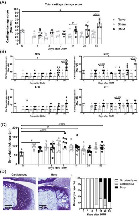
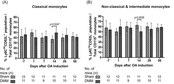
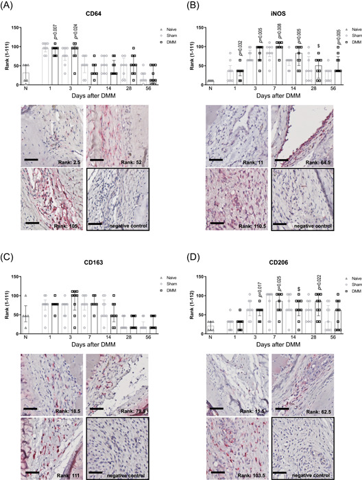
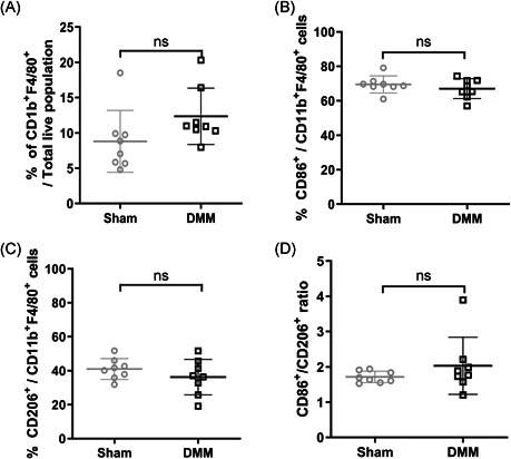
Macrophages play an important role in the development and progression of osteoarthritis (OA). The aim of this study was to identify macrophage phenotypes in synovium and monocyte subsets in peripheral blood in C57BL/6 mice by destabilizing the medial meniscus (DMM), and the association of macrophage subsets with OA features. DMM, sham, and non-operated knees were histologically assessed between 1 and 56 days for macrophage polarization states by immunohistochemistry (IHC), cartilage damage, synovial thickening, and osteophytes (n = 9 per timepoint). Naive knees (n = 6) were used as controls. Monocyte and polarized synovial macrophage subsets were evaluated by flow cytometry. CD64 and CD206 levels on IHC were higher at early timepoints in DMM and sham knees compared to naive knees. iNOS labeling intensity was higher in DMM and sham knees than in naive knees from d3 onwards. CD163 expression was unaltered at all timepoints. Even though macrophage polarization profiles were similar in DMM and sham knees, only in DMM knees the presence of iNOS and CD206 associated with synovial thickness, and CD163 staining inversely correlated with osteophyte presence. At day 14, monocyte subset distribution was different in peripheral blood of DMM mice compared with sham mice. In conclusion, monocyte subsets in blood and synovial macrophage phenotypes vary after joint surgery. High levels of iNOS , CD163 , and CD206 cells are found in both destabilized and sham-operated knees, and coexistence with joint instability may be a requirement to initiate and exacerbate OA progression.
巨噬细胞在骨关节炎(OA)的发展和进程中发挥着重要作用。本研究的目的是通过破坏内侧半月板(DMM)来确定C57BL/6小鼠滑膜中的巨噬细胞表型和外周血中的单核细胞亚群,以及巨噬细胞亚群与OA特征之间的关联。在1至56天之间,通过免疫组织化学(IHC)对DMM组、假手术组和未手术组的膝关节进行组织学评估,以确定巨噬细胞极化状态、软骨损伤、滑膜增厚和骨赘形成情况(每个时间点n = 9)。将未处理的膝关节(n = 6)用作对照。通过流式细胞术评估单核细胞和极化的滑膜巨噬细胞亚群。与未处理的膝关节相比,DMM组和假手术组膝关节在早期时间点的IHC上CD64和CD206水平更高。从第3天起,DMM组和假手术组膝关节中的诱导型一氧化氮合酶(iNOS)标记强度高于未处理的膝关节。在所有时间点,CD163表达均未改变。尽管DMM组和假手术组膝关节中的巨噬细胞极化谱相似,但仅在DMM组膝关节中,iNOS和CD206的存在与滑膜厚度相关,而CD163染色与骨赘的存在呈负相关。在第14天,DMM小鼠外周血中的单核细胞亚群分布与假手术小鼠不同。总之,关节手术后血液中的单核细胞亚群和滑膜巨噬细胞表型会发生变化。在DMM组和假手术组膝关节中均发现高水平的iNOS、CD163和CD206细胞,与关节不稳定共存可能是启动和加剧OA进展的必要条件。