Experimental Rheumatology, Department of Rheumatology, Radboud University Medical Center, PO Box 9101, Nijmegen, 6500 HB, The Netherlands.
Institute of Immunology, University of Munster, Munster, Germany.
Arthritis Res Ther. 2017 Sep 29;19(1):217. doi: 10.1186/s13075-017-1426-6.
Monocytes are dominant cells present within the inflamed synovium during osteoarthritis (OA). In mice, two functionally distinct monocyte subsets are described: pro-inflammatory Ly6C and patrolling Ly6C monocytes. Alarmins S100A8/A9 locally released by the synovium during inflammatory OA for prolonged periods may be dominant proteins involved in stimulating recruitment of Ly6C monocytes from the circulation to the joint. Our objective was to investigate the role of S100A8/A9 in the mobilization of Ly6C and Ly6C monocytic populations to the inflamed joint in collagenase-induced OA (CiOA).
S100A8 was injected intra-articularly to investigate monocyte influx. CiOA was induced by injection of collagenase into knee joints of wild-type C57BL/6 (WT), and S100a9 mice. Mice were sacrificed together with age-matched saline-injected control mice (n = 6/group), and expression of monocyte markers, pro-inflammatory cytokines, and chemokines was determined in the synovium using ELISA and RT-qPCR. Cells were isolated from the bone marrow (BM), spleen, blood, and synovium and monocytes were identified using FACS.
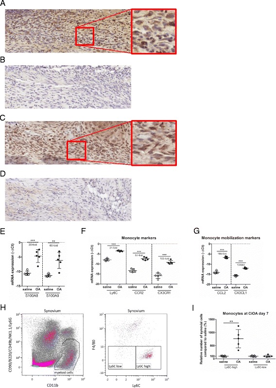
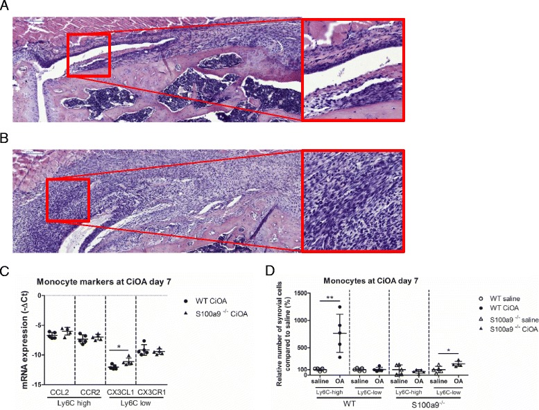
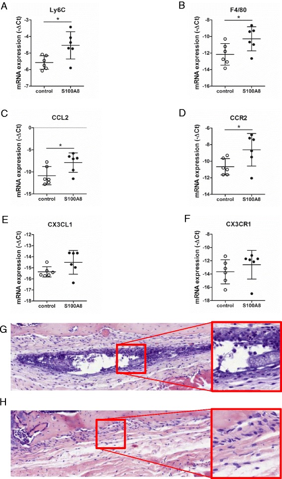
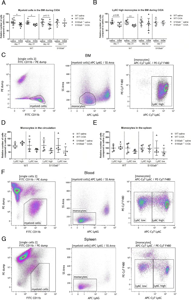
S100A8/A9 was highly expressed during CiOA. Intra-articular injection of S100A8 leads to elevated expression of monocyte markers and the monocyte-attracting chemokines CCL2 and CX3CL1 in the synovium. At day 7 (d7) after CiOA induction in WT mice, numbers of Ly6C, but not Ly6C monocytes, were strongly increased (7.6-fold) in the synovium compared to saline-injected controls. This coincided with strong upregulation of CCL2, which preferentially attracts Ly6C monocytes. In contrast, S100a9 mice showed a significant increase in Ly6C monocytes (twofold) within the synovium at CiOA d7, whereas the number of Ly6C monocytes remained unaffected. In agreement with this finding, the Ly6C mobilization marker CX3CL1 was significantly higher within the synovium of S100a9 mice. Next, we studied the effect of S100A8/A9 on release of Ly6C monocytes from the BM into the circulation. A 14% decrease in myeloid cells was found in WT BM at CiOA d7. No decrease in myeloid cells in S100a9 BM was found, suggesting that S100A8/A9 promotes the release of myeloid populations from the BM.
Induction of OA locally leads to strongly elevated S100A8/A9 expression and an elevated influx of Ly6C monocytes from the BM to the synovium.
在骨关节炎(OA)期间,单核细胞是炎症滑膜中占优势的细胞。在小鼠中,描述了两种功能上不同的单核细胞亚群:促炎 Ly6C 和巡逻 Ly6C 单核细胞。在炎症性 OA 期间,滑膜局部持续释放的警报素 S100A8/A9 可能是刺激 Ly6C 单核细胞从循环募集到关节的主要蛋白。我们的目的是研究 S100A8/A9 在胶原酶诱导的 OA(CiOA)中动员 Ly6C 和 Ly6C 单核细胞群进入炎症关节中的作用。
关节内注射 S100A8 以研究单核细胞浸润。通过向野生型 C57BL/6(WT)和 S100a9 小鼠的膝关节内注射胶原酶来诱导 CiOA。与年龄匹配的盐水注射对照小鼠(每组 n=6)一起处死小鼠,并使用 ELISA 和 RT-qPCR 确定滑膜中单核细胞标志物、促炎细胞因子和趋化因子的表达。从骨髓(BM)、脾脏、血液和滑膜中分离细胞,并使用 FACS 鉴定单核细胞。
S100A8/A9 在 CiOA 期间高度表达。关节内注射 S100A8 导致滑膜中单核细胞标志物和单核细胞趋化因子 CCL2 和 CX3CL1 的表达升高。在 CiOA 诱导后第 7 天(d7),与盐水注射对照相比,WT 小鼠的滑膜中 Ly6C 但不是 Ly6C 单核细胞的数量强烈增加(7.6 倍)。这与 CCL2 的强烈上调一致,CCL2 优先吸引 Ly6C 单核细胞。相比之下,S100a9 小鼠在 CiOA d7 时滑膜中 Ly6C 单核细胞数量显著增加(两倍),而 Ly6C 单核细胞数量不受影响。与这一发现一致的是,Ly6C 动员标志物 CX3CL1 在 S100a9 小鼠的滑膜中显著升高。接下来,我们研究了 S100A8/A9 对骨髓中 Ly6C 单核细胞释放到循环中的影响。在 CiOA d7 时,WT BM 中的髓样细胞减少了 14%。在 S100a9 BM 中未发现髓样细胞减少,表明 S100A8/A9 促进骨髓中髓样细胞的释放。
OA 的诱导导致局部 S100A8/A9 表达的强烈升高和 Ly6C 单核细胞从骨髓向滑膜的流入增加。