Department of Biomedical Imaging and Radiological Sciences, National Yang-Ming University, Taipei, Taiwan.
Department of Surgery, Division of Thoracic Surgery, MacKay Memorial Hospital, Taipei, Taiwan.
Aging Cell. 2021 Jan;20(1):e13288. doi: 10.1111/acel.13288. Epub 2020 Dec 18.
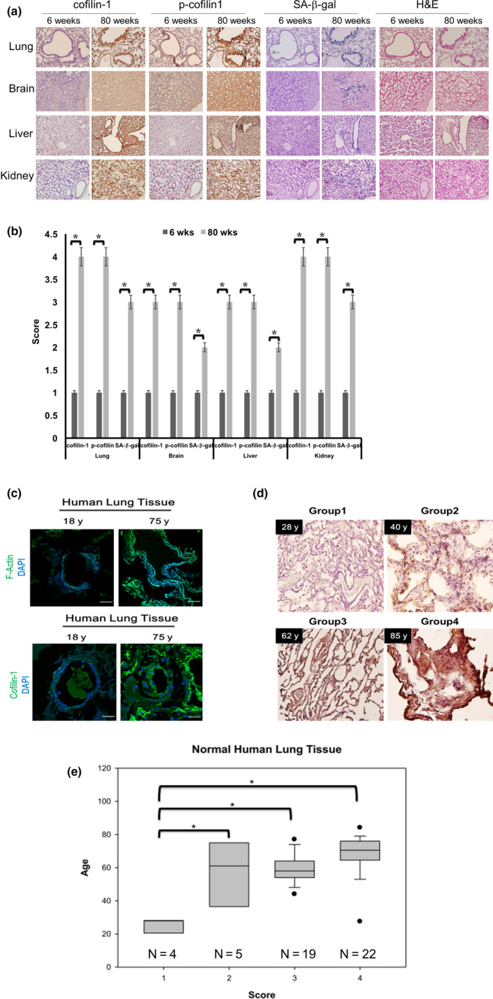
Morphological change is an explicit characteristic of cell senescence, but the underlying mechanisms remains to be addressed. Here, we demonstrated, after a survey of various actin-binding proteins, that the post-translational up-regulation of cofilin-1 was essential for the reduced rate of actin depolymerization morphological enlargement in senescent cells. Additionally, up-regulated cofilin-1 mainly existed in the serine-3 phosphorylated form, according to the 2D gel immunoblotting assay. The up-regulation of cofilin-1 was also detected in aged mammalian tissues. The over-expression of wild-type cofilin-1 and constitutively phosphorylated cofilin-1 promoted cell senescence with an increased cell size. Additionally, senescent phenotypes were also reduced by knockdown of total cofilin-1, which led to a decrease in phosphorylated cofilin-1. The senescence induced by the over-expression of cofilin-1 was dependent on p27 , but not on the p53 and p16 expressions. The knockdown of p27 alleviated cell senescence induced by oxidative stress or replicative stress. We also found that the over-expression of cofilin-1 induced the expression of p27 through transcriptional suppression of the transcriptional enhancer factors domain 1 (TEAD1) transcription factor. The TEAD1 transcription factor played a transrepressive role in the p27 gene promoter, as determined by the promoter deletion reporter gene assay. Interestingly, the down-regulation of TEAD1 was accompanied by the up-regulation of cofilin-1 in senescence. The knockdown and restoration of TEAD1 in young cells and old cells could induce and inhibit p27 and senescent phenotypes, respectively. Taken together, the current data suggest that cofilin-1/TEAD1/p27 signaling is involved in senescence-related morphological change and growth arrest.
形态改变是细胞衰老的一个明显特征,但其中的机制仍有待阐明。在这里,我们在调查了各种肌动蛋白结合蛋白后发现,在后翻译水平上,上调原肌球蛋白 1(cofilin-1)对于衰老细胞中肌动蛋白解聚率降低导致的形态增大至关重要。此外,根据二维凝胶免疫印迹分析,上调的 cofilin-1 主要以丝氨酸 3 磷酸化形式存在。在衰老的哺乳动物组织中也检测到了 cofilin-1 的上调。野生型 cofilin-1 和组成型磷酸化 cofilin-1 的过表达促进了细胞衰老,并使细胞体积增大。此外,敲低总 cofilin-1 也减少了衰老表型,导致磷酸化 cofilin-1 减少。cofilin-1 的过表达诱导的衰老依赖于 p27,但不依赖于 p53 和 p16 的表达。p27 的敲低减轻了由氧化应激或复制应激引起的细胞衰老。我们还发现,cofilin-1 的过表达通过转录抑制转录增强因子域 1(TEAD1)转录因子诱导 p27 的表达。TEAD1 转录因子在 p27 基因启动子上发挥反式抑制作用,如启动子缺失报告基因分析所示。有趣的是,在衰老过程中,TEAD1 的下调伴随着 cofilin-1 的上调。在年轻细胞和衰老细胞中敲低和恢复 TEAD1 可以分别诱导和抑制 p27 和衰老表型。总之,目前的数据表明,cofilin-1/TEAD1/p27 信号通路参与了与衰老相关的形态变化和生长停滞。