• 文献检索
  • 文档翻译
  • 深度研究
  • 学术资讯
  • Suppr Zotero 插件Zotero 插件
  • 邀请有礼
  • 套餐&价格
  • 历史记录
应用&插件
Suppr Zotero 插件Zotero 插件浏览器插件Mac 客户端Windows 客户端微信小程序
定价
高级版会员购买积分包购买API积分包
服务
文献检索文档翻译深度研究API 文档MCP 服务
关于我们
关于 Suppr公司介绍联系我们用户协议隐私条款
关注我们

Suppr 超能文献

核心技术专利:CN118964589B侵权必究
粤ICP备2023148730 号-1Suppr @ 2026

文献检索

告别复杂PubMed语法,用中文像聊天一样搜索,搜遍4000万医学文献。AI智能推荐,让科研检索更轻松。

立即免费搜索

文件翻译

保留排版,准确专业,支持PDF/Word/PPT等文件格式,支持 12+语言互译。

免费翻译文档

深度研究

AI帮你快速写综述,25分钟生成高质量综述,智能提取关键信息,辅助科研写作。

立即免费体验

胶质母细胞瘤术后瘤周水肿区较高的Cho/NAA比值与较早复发相关。

Higher Cho/NAA Ratio in Postoperative Peritumoral Edema Zone Is Associated With Earlier Recurrence of Glioblastoma.

作者信息

Cui Yong, Zeng Wei, Jiang Haihui, Ren Xiaohui, Lin Song, Fan Yanzhu, Liu Yapeng, Zhao Jizong

机构信息

Department of Neurosurgery, Beijing Tiantan Hospital, Capital Medical University, Beijing, China.

National Clinical Research Center for Neurological Diseases, Center of Brain Tumor, Beijing Institute for Brain Disorders and Beijing Key Laboratory of Brain Tumor, Beijing, China.

出版信息

Front Neurol. 2020 Dec 4;11:592155. doi: 10.3389/fneur.2020.592155. eCollection 2020.

DOI:10.3389/fneur.2020.592155
PMID:33343496
原文链接:https://pmc.ncbi.nlm.nih.gov/articles/PMC7747764/
Abstract

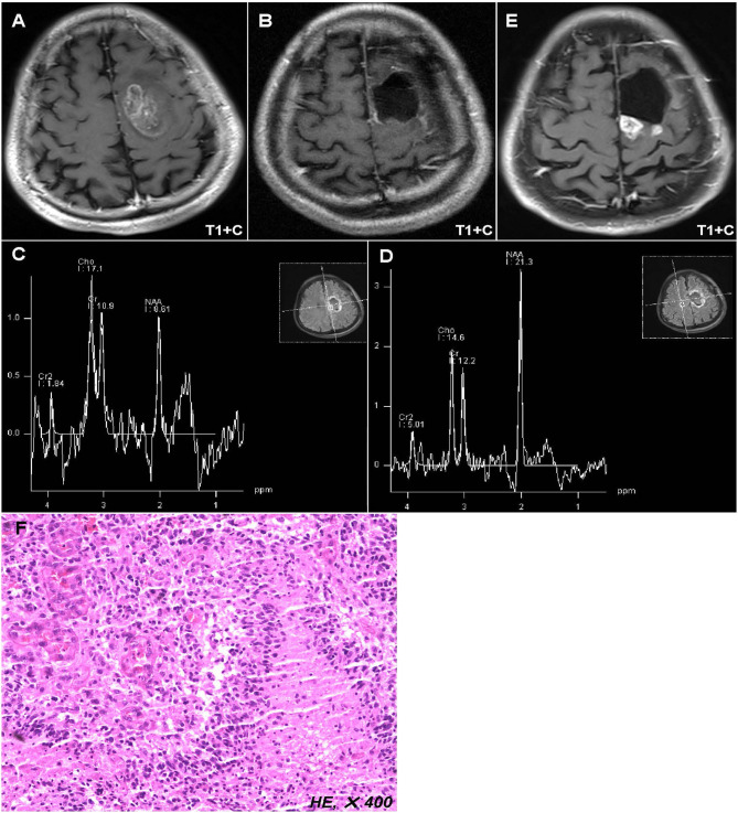
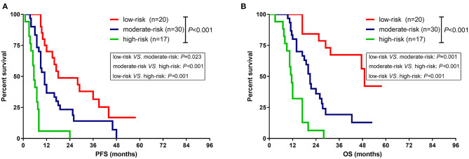
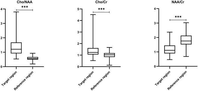
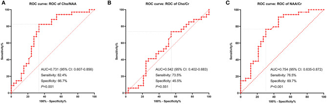
To explore the prognostic significance of metabolic parameters in postoperative peritumoral edema zone (PEZ) of patients with glioblastoma (GBM) based on proton magnetic resonance spectroscopy (MRS). The postoperative MRS data of 67 patients with GBM from Beijing Tiantan Hospital were retrospectively reviewed. Metabolite ratios including Cho/NAA, Cho/Cr, and NAA/Cr in both postoperative PEZ and contralateral normal brain region were recorded. Log-rank analysis and Cox regression model were used to identify parameters correlated with progression-free survival (PFS) and overall survival (OS). Compared with the contralateral normal brain region, postoperative PEZ showed a lower ratio of NAA/Cr (1.20 ± 0.42 vs. 1.81 ± 0.48, < 0.001), and higher ratios of Cho/Cr and Cho/NAA (1.36 ± 0.44 vs. 1.02 ± 0.27, < 0.001 and 1.32 ± 0.59 vs. 0.57 ± 0.14, < 0.001). Both the ratios of Cho/NAA and NAA/Cr were identified as prognostic factors in univariate analysis ( < 0.05), while only Cho/NAA ≥ 1.31 was further confirmed as an independent risk factor for early recurrence in the Cox regression model ( < 0.01). According to the factors of MGMT promoter unmethylation, without radiotherapy and Cho/NAA ≥ 1.31, a prognostic scoring scale for GBM was established, which could divide patients into low-risk, moderate-risk, and high-risk groups. There was a significant difference of survival rate between the three groups ( < 0.001). Higher Cho/NAA ratio in the postoperative PEZ of GBM predicts earlier recurrence and is associated with poor prognosis. The prognostic scoring scale based on clinical, molecular and metabolic parameters of patients with GBM can help doctors to make more precise prediction of survival time and to adjust therapeutic regimens.

摘要

基于质子磁共振波谱(MRS)探讨胶质母细胞瘤(GBM)患者术后瘤周水肿区(PEZ)代谢参数的预后意义。回顾性分析北京天坛医院67例GBM患者的术后MRS数据。记录术后PEZ及对侧正常脑区的代谢物比率,包括胆碱/ N-乙酰天门冬氨酸(Cho/NAA)、胆碱/肌酸(Cho/Cr)和N-乙酰天门冬氨酸/肌酸(NAA/Cr)。采用对数秩分析和Cox回归模型确定与无进展生存期(PFS)和总生存期(OS)相关的参数。与对侧正常脑区相比,术后PEZ的NAA/Cr比率较低(1.20±0.42 vs. 1.81±0.48,<0.001),Cho/Cr和Cho/NAA比率较高(1.36±0.44 vs. 1.02±0.27,<0.001;1.32±0.59 vs. 0.57±0.14,<0.001)。单因素分析中,Cho/NAA和NAA/Cr比率均被确定为预后因素(<0.05),而在Cox回归模型中,只有Cho/NAA≥1.31被进一步确认为早期复发的独立危险因素(<0.01)。根据O6-甲基鸟嘌呤-DNA甲基转移酶(MGMT)启动子未甲基化、未接受放疗和Cho/NAA≥1.31等因素,建立了GBM预后评分量表,可将患者分为低风险、中风险和高风险组。三组间生存率有显著差异(<0.001)。GBM术后PEZ中较高的Cho/NAA比率预示着更早复发,并与预后不良相关。基于GBM患者临床、分子和代谢参数的预后评分量表有助于医生更精确地预测生存时间并调整治疗方案。

https://cdn.ncbi.nlm.nih.gov/pmc/blobs/403c/7747764/041013e55b41/fneur-11-592155-g0004.jpg
https://cdn.ncbi.nlm.nih.gov/pmc/blobs/403c/7747764/04a2dc11b549/fneur-11-592155-g0001.jpg
https://cdn.ncbi.nlm.nih.gov/pmc/blobs/403c/7747764/02e2d2a59096/fneur-11-592155-g0002.jpg
https://cdn.ncbi.nlm.nih.gov/pmc/blobs/403c/7747764/e58d50354b08/fneur-11-592155-g0003.jpg
https://cdn.ncbi.nlm.nih.gov/pmc/blobs/403c/7747764/041013e55b41/fneur-11-592155-g0004.jpg

相似文献

1
Higher Cho/NAA Ratio in Postoperative Peritumoral Edema Zone Is Associated With Earlier Recurrence of Glioblastoma.胶质母细胞瘤术后瘤周水肿区较高的Cho/NAA比值与较早复发相关。
Front Neurol. 2020 Dec 4;11:592155. doi: 10.3389/fneur.2020.592155. eCollection 2020.
2
Major Metabolite Levels of Preoperative Proton Magnetic Resonance Sectroscopy and Intraoperative Fluorescence Intensity in Glioblastoma.胶质母细胞瘤术前质子磁共振波谱主要代谢物水平与术中荧光强度
Zhongguo Yi Xue Ke Xue Yuan Xue Bao. 2017 Aug 20;39(4):511-517. doi: 10.3881/j.issn.1000-503X.2017.04.009.
3
Proton magnetic resonance spectroscopy in the distinction of high-grade cerebral gliomas from single metastatic brain tumors.质子磁共振波谱在鉴别高级别脑胶质瘤与单发性脑转移瘤中的应用
Acta Radiol. 2010 Apr;51(3):316-25. doi: 10.3109/02841850903482901.
4
ACRIN 6684: Multicenter, phase II assessment of tumor hypoxia in newly diagnosed glioblastoma using magnetic resonance spectroscopy.ACRIN 6684:采用磁共振波谱技术评估新诊断胶质母细胞瘤肿瘤缺氧的多中心、二期研究。
PLoS One. 2018 Jun 14;13(6):e0198548. doi: 10.1371/journal.pone.0198548. eCollection 2018.
5
Correlation analysis of expressions of PTEN and p53 with the value obtained by magnetic resonance spectroscopy and apparent diffusion coefficient in the tumor and the tumor-adjacent area in magnetic resonance imaging for glioblastoma.胶质母细胞瘤中,肿瘤及肿瘤周边区域磁共振波谱值和表观扩散系数与PTEN和p53表达的相关性分析(磁共振成像)
J BUON. 2018 Mar-Apr;23(2):391-397.
6
Brain Metastases of Non-Small Cell Lung Cancer: Magnetic Resonance Spectroscopy for Clinical Outcome Assessment in Patients with Stereotactic Radiotherapy.非小细胞肺癌脑转移:立体定向放射治疗患者临床结局评估的磁共振波谱分析
Onco Targets Ther. 2020 Dec 22;13:13087-13096. doi: 10.2147/OTT.S286893. eCollection 2020.
7
Measurements of diagnostic examination performance using quantitative apparent diffusion coefficient and proton MR spectroscopic imaging in the preoperative evaluation of tumor grade in cerebral gliomas.使用定量表观扩散系数和质子磁共振波谱成像对脑胶质瘤术前肿瘤分级进行诊断检查性能的测量。
Eur J Radiol. 2011 Nov;80(2):462-70. doi: 10.1016/j.ejrad.2010.07.017. Epub 2010 Aug 13.
8
[Value of proton magnetic resonance spectroscopy with two-dimensional chemical-shift imaging in evaluating brain gliomas].二维化学位移成像质子磁共振波谱在评估脑胶质瘤中的价值
Nan Fang Yi Ke Da Xue Xue Bao. 2008 Aug;28(8):1342-4.
9
MR spectroscopy using normalized and non-normalized metabolite ratios for differentiating recurrent brain tumor from radiation injury.利用归一化和非归一化代谢物比值的磁共振波谱分析鉴别脑肿瘤复发与放射性损伤。
Acad Radiol. 2011 Sep;18(9):1101-8. doi: 10.1016/j.acra.2011.05.006.
10
The diagnostic performance of magnetic resonance spectroscopy in differentiating high-from low-grade gliomas: A systematic review and meta-analysis.磁共振波谱在鉴别高级别与低级别胶质瘤中的诊断性能:一项系统评价和荟萃分析。
Eur Radiol. 2016 Aug;26(8):2670-84. doi: 10.1007/s00330-015-4046-z. Epub 2015 Oct 15.

引用本文的文献

1
Glioblastoma metabolomics: uncovering biomarkers for diagnosis, prognosis and targeted therapy.胶质母细胞瘤代谢组学:揭示用于诊断、预后和靶向治疗的生物标志物。
J Exp Clin Cancer Res. 2025 Aug 7;44(1):230. doi: 10.1186/s13046-025-03497-2.
2
Defining occult disease in glioblastoma using spectroscopic MRI: implications for clinical target volume delineation.利用磁共振波谱成像定义胶质母细胞瘤中的隐匿性疾病:对临床靶区勾画的意义
Radiat Oncol. 2025 May 22;20(1):86. doi: 10.1186/s13014-025-02666-z.
3
Impact of Sex Hormones on Glioblastoma: Sex-Related Differences and Neuroradiological Insights.

本文引用的文献

1
CBTRUS Statistical Report: Primary Brain and Other Central Nervous System Tumors Diagnosed in the United States in 2012-2016.美国 2012-2016 年诊断的原发性脑和其他中枢神经系统肿瘤 CBTRUS 统计报告。
Neuro Oncol. 2019 Nov 1;21(Suppl 5):v1-v100. doi: 10.1093/neuonc/noz150.
2
Multimodal MRI characteristics of the glioblastoma infiltration beyond contrast enhancement.胶质母细胞瘤对比增强以外浸润的多模态磁共振成像特征
Ther Adv Neurol Disord. 2019 May 14;12:1756286419844664. doi: 10.1177/1756286419844664. eCollection 2019.
3
Assessment of Overall Survival in Glioma Patients as Predicted by Metabolomic Criteria.
性激素对胶质母细胞瘤的影响:性别差异与神经放射学见解
Life (Basel). 2024 Nov 21;14(12):1523. doi: 10.3390/life14121523.
4
Turning attention to tumor-host interface and focus on the peritumoral heterogeneity of glioblastoma.将注意力转向肿瘤-宿主界面,并聚焦于胶质母细胞瘤的瘤周异质性。
Nat Commun. 2024 Dec 30;15(1):10885. doi: 10.1038/s41467-024-55243-5.
5
Peritumoral Brain Zone in Astrocytoma: Morphology, Molecular Aspects, and Clinical Manifestations (Review).脑胶质瘤瘤周区:形态学、分子学方面和临床表现(综述)。
Sovrem Tekhnologii Med. 2024;16(2):79-88. doi: 10.17691/stm2024.16.2.08. Epub 2024 Apr 27.
6
Improved Recovery of Complete Spinal Cord Transection by a Plasma-Modified Fibrillar Scaffold.通过等离子体改性纤维支架改善完全性脊髓横断的恢复情况。
Polymers (Basel). 2024 Apr 18;16(8):1133. doi: 10.3390/polym16081133.
7
Peritumoral brain zone in glioblastoma: biological, clinical and mechanical features.胶质母细胞瘤瘤周脑区:生物学、临床和力学特征。
Front Immunol. 2024 Feb 29;15:1347877. doi: 10.3389/fimmu.2024.1347877. eCollection 2024.
8
The STEMRI trial: Magnetic resonance spectroscopy imaging can define tumor areas enriched in glioblastoma stem-like cells.STEMRI 试验:磁共振波谱成像可界定富含胶质母细胞瘤干细胞样细胞的肿瘤区域。
Sci Adv. 2023 Nov 3;9(44):eadi0114. doi: 10.1126/sciadv.adi0114.
9
Advanced Magnetic Resonance Imaging in the Evaluation of Treated Glioblastoma: A Pictorial Essay.高级磁共振成像在治疗后胶质母细胞瘤评估中的应用:图文综述
Cancers (Basel). 2023 Jul 26;15(15):3790. doi: 10.3390/cancers15153790.
10
Conventional and Advanced Magnetic Resonance Imaging Assessment of Non-Enhancing Peritumoral Area in Brain Tumor.脑肿瘤中无强化瘤周区域的传统及高级磁共振成像评估
Cancers (Basel). 2023 May 30;15(11):2992. doi: 10.3390/cancers15112992.
根据代谢组学标准预测的胶质瘤患者总生存期评估
Front Oncol. 2019 May 10;9:328. doi: 10.3389/fonc.2019.00328. eCollection 2019.
4
Proliferation-dominant high-grade astrocytoma: survival benefit associated with extensive resection of FLAIR abnormality region.增殖为主型高级别星形细胞瘤:与FLAIR异常区域广泛切除相关的生存获益
J Neurosurg. 2019 Mar 22;132(4):998-1005. doi: 10.3171/2018.12.JNS182775. Print 2020 Apr 1.
5
Adult Glioblastoma.成人胶质母细胞瘤。
J Clin Oncol. 2017 Jul 20;35(21):2402-2409. doi: 10.1200/JCO.2017.73.0119. Epub 2017 Jun 22.
6
Quantitative MRI for analysis of peritumoral edema in malignant gliomas.用于分析恶性胶质瘤瘤周水肿的定量磁共振成像
PLoS One. 2017 May 23;12(5):e0177135. doi: 10.1371/journal.pone.0177135. eCollection 2017.
7
Pathological analysis of the surgical margins of resected glioblastomas excised using photodynamic visualization with both 5-aminolevulinic acid and fluorescein sodium.使用5-氨基乙酰丙酸和荧光素钠进行光动力可视化切除的胶质母细胞瘤手术切缘的病理分析。
J Neurooncol. 2017 Jun;133(2):389-397. doi: 10.1007/s11060-017-2445-5. Epub 2017 Apr 21.
8
Dual-labeling with 5-aminolevulinic acid and fluorescein for fluorescence-guided resection of high-grade gliomas: technical note.5-氨基酮戊酸和荧光素双重标记在高级别胶质瘤荧光引导切除术中的应用:技术说明。
J Neurosurg. 2018 Feb;128(2):399-405. doi: 10.3171/2016.11.JNS161072. Epub 2017 Mar 24.
9
Association of early changes in 1H MRSI parameters with survival for patients with newly diagnosed glioblastoma receiving a multimodality treatment regimen.接受多模态治疗方案的新诊断胶质母细胞瘤患者1H磁共振波谱成像(MRSI)参数早期变化与生存的相关性
Neuro Oncol. 2017 Mar 1;19(3):430-439. doi: 10.1093/neuonc/now159.
10
Association of the Extent of Resection With Survival in Glioblastoma: A Systematic Review and Meta-analysis.胶质母细胞瘤切除范围与生存的相关性:一项系统评价和荟萃分析。
JAMA Oncol. 2016 Nov 1;2(11):1460-1469. doi: 10.1001/jamaoncol.2016.1373.