Yan Jiun-Lin, Li Chao, Boonzaier Natalie R, Fountain Daniel M, Larkin Timothy J, Matys Tomasz, van der Hoorn Anouk, Price Stephen J
Department of Neurosurgery, Chang Gung University College of Medicine, Taoyuan, Taiwan.
Cambridge Brain Tumour Imaging Laboratory, Division of Neurosurgery and Wolfson Brain Imaging Center, Department of Clinical Neuroscience, University of Cambridge, Addenbrooke's Hospital, Box 167, CB2 0QQ, Cambridge, UK.
Ther Adv Neurol Disord. 2019 May 14;12:1756286419844664. doi: 10.1177/1756286419844664. eCollection 2019.
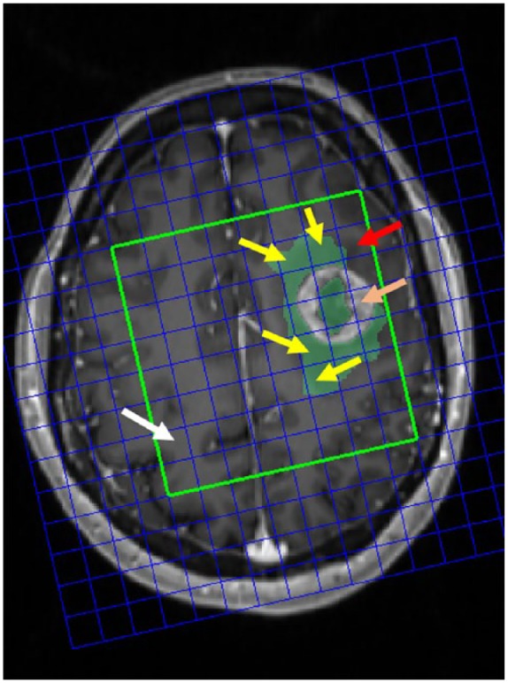
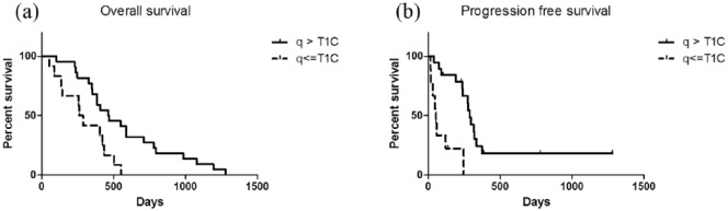
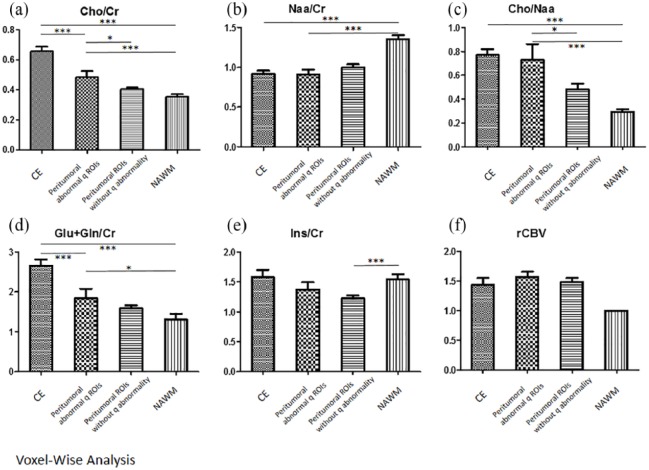
Our inability to identify the invasive margin of glioblastomas hampers attempts to achieve local control. Diffusion tensor imaging (DTI) has been implemented clinically to delineate the margin of the tumor infiltration, its derived anisotropic (q) values can extend beyond the contrast-enhanced area and correlates closely with the tumor. However, its correlation with tumor infiltration shown on multivoxel proton magnetic resonance spectroscopy (MRS) and perfusion magnetic resonance imaging (MRI) should be investigated. In this study, we aimed to show tissue characteristics of the q-defined peritumoral invasion on MRS and perfusion MRI. Patients with a primary glioblastoma were included ( = 51). Four regions of interest were analyzed; the contrast-enhanced lesion, peritumoral abnormal q region, peritumoral normal q region, and contralateral normal-appearing white matter. MRS, including choline (Cho)/creatinine (Cr), Cho/N-acetyl-aspartate (NAA) and NAA/Cr ratios, and the relative cerebral blood volume (rCBV) were analyzed. Our results showed an increase in the Cho/NAA ( = 0.0346) and Cho/Cr ( = 0.0219) ratios in the peritumoral abnormal q region, suggestive of tumor invasion. The rCBV was marginally elevated ( = 0.0798). Furthermore, the size of the abnormal q regions was correlated with survival; patients with larger abnormal q regions showed better progression-free survival (median 287 53 days, = 0.001) and overall survival (median 464 274 days, = 0.006) than those with smaller peritumoral abnormal q regions of interest. These results support how the DTI q abnormal area identifies tumor activity beyond the contrast-enhanced area, especially correlating with MRS.
我们无法确定胶质母细胞瘤的浸润边缘,这阻碍了实现局部控制的努力。扩散张量成像(DTI)已在临床上用于描绘肿瘤浸润边缘,其衍生的各向异性(q)值可延伸至增强区域之外,并与肿瘤密切相关。然而,应研究其与多体素质子磁共振波谱(MRS)和灌注磁共振成像(MRI)所示肿瘤浸润的相关性。在本研究中,我们旨在展示MRS和灌注MRI上q定义的瘤周浸润的组织特征。纳入原发性胶质母细胞瘤患者(n = 51)。分析了四个感兴趣区域;增强病变、瘤周异常q区域、瘤周正常q区域和对侧外观正常的白质。分析了MRS,包括胆碱(Cho)/肌酐(Cr)、Cho/N-乙酰天门冬氨酸(NAA)和NAA/Cr比值,以及相对脑血容量(rCBV)。我们的结果显示,瘤周异常q区域的Cho/NAA(P = 0.0346)和Cho/Cr(P = 0.0219)比值增加,表示肿瘤浸润。rCBV略有升高(P = 0.0798)。此外,异常q区域的大小与生存率相关;与瘤周异常q感兴趣区域较小的患者相比,异常q区域较大的患者无进展生存期更好(中位数287±53天,P = 0.001),总生存期更好(中位数464±274天,P = 0.006)。这些结果支持了DTI的q异常区域如何识别增强区域之外的肿瘤活性,尤其是与MRS相关。