• 文献检索
  • 文档翻译
  • 深度研究
  • 学术资讯
  • Suppr Zotero 插件Zotero 插件
  • 邀请有礼
  • 套餐&价格
  • 历史记录
应用&插件
Suppr Zotero 插件Zotero 插件浏览器插件Mac 客户端Windows 客户端微信小程序
定价
高级版会员购买积分包购买API积分包
服务
文献检索文档翻译深度研究API 文档MCP 服务
关于我们
关于 Suppr公司介绍联系我们用户协议隐私条款
关注我们

Suppr 超能文献

核心技术专利:CN118964589B侵权必究
粤ICP备2023148730 号-1Suppr @ 2026

文献检索

告别复杂PubMed语法,用中文像聊天一样搜索,搜遍4000万医学文献。AI智能推荐,让科研检索更轻松。

立即免费搜索

文件翻译

保留排版,准确专业,支持PDF/Word/PPT等文件格式,支持 12+语言互译。

免费翻译文档

深度研究

AI帮你快速写综述,25分钟生成高质量综述,智能提取关键信息,辅助科研写作。

立即免费体验

用于对导管原位癌进行分级的核异质性图像指数的推导

Derivation of a nuclear heterogeneity image index to grade DCIS.

作者信息

Hayward Mary-Kate, Louise Jones J, Hall Allison, King Lorraine, Ironside Alastair J, Nelson Andrew C, Shelley Hwang E, Weaver Valerie M

机构信息

Center for Bioengineering and Tissue Regeneration, Department of Surgery, University of California San Francisco, San Francisco, CA, USA.

Center for Tumor Biology, Barts Cancer Institute, John Vane Science Building, Barts and the London School of Medicine and Dentistry, UK.

出版信息

Comput Struct Biotechnol J. 2020 Dec 3;18:4063-4070. doi: 10.1016/j.csbj.2020.11.040. eCollection 2020.

DOI:10.1016/j.csbj.2020.11.040
PMID:33363702
原文链接:https://pmc.ncbi.nlm.nih.gov/articles/PMC7744935/
Abstract

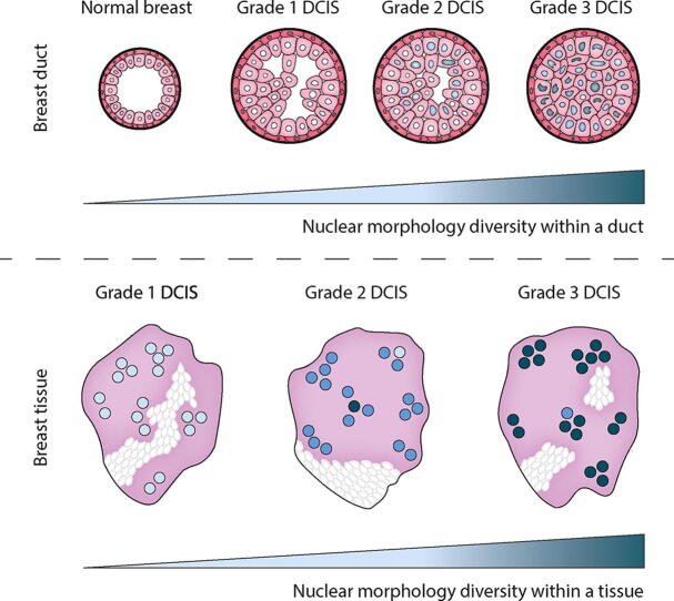
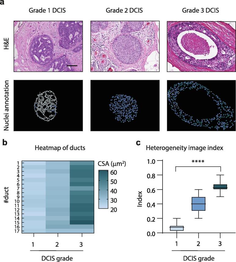
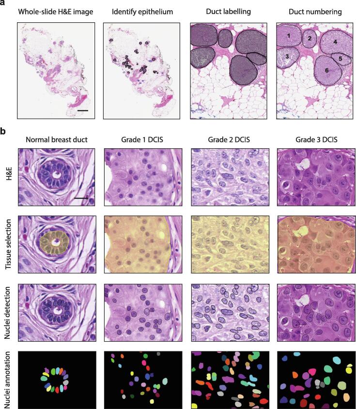
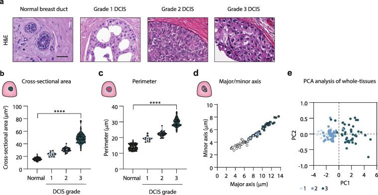
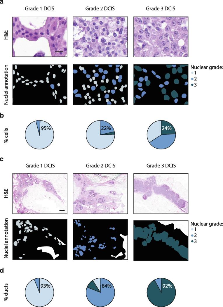
Abnormalities in cell nuclear morphology are a hallmark of cancer. Histological assessment of cell nuclear morphology is frequently used by pathologists to grade ductal carcinoma in situ (DCIS). Objective methods that allow standardization and reproducibility of cell nuclear morphology assessment have potential to improve the criteria needed to predict DCIS progression and recurrence. Aggressive cancers are highly heterogeneous. We asked whether cell nuclear morphology heterogeneity could be incorporated into a metric to classify DCIS. We developed a nuclear heterogeneity image index to objectively, and quantitatively grade DCIS. A whole-tissue cell nuclear morphological analysis, that classified tumors by the worst ten percent in a duct-by-duct manner, identified nuclear size ranges associated with each DCIS grade. Digital image analysis further revealed increasing heterogeneity within ducts or between ducts in tissues of worsening DCIS grade. The findings illustrate how digital image analysis comprises a supplemental tool for pathologists to objectively classify DCIS and in the future, may provide a method to predict patient outcome through analysis of nuclear heterogeneity.

摘要

细胞核形态异常是癌症的一个标志。病理学家经常使用细胞核形态的组织学评估来对导管原位癌(DCIS)进行分级。能够实现细胞核形态评估标准化和可重复性的客观方法有潜力改进预测DCIS进展和复发所需的标准。侵袭性癌症具有高度异质性。我们询问细胞核形态异质性是否可以纳入一个指标来对DCIS进行分类。我们开发了一种核异质性图像指数来客观、定量地对DCIS进行分级。一种全组织细胞核形态分析,以逐导管方式根据最差的百分之十对肿瘤进行分类,确定了与每个DCIS分级相关的核大小范围。数字图像分析进一步揭示,在DCIS分级恶化的组织中,导管内或导管间的异质性在增加。这些发现说明了数字图像分析如何构成一种辅助工具,供病理学家客观地对DCIS进行分类,并且在未来,可能通过分析核异质性提供一种预测患者预后的方法。

https://cdn.ncbi.nlm.nih.gov/pmc/blobs/1c01/7744935/4566e5718d16/gr4.jpg
https://cdn.ncbi.nlm.nih.gov/pmc/blobs/1c01/7744935/43595ee08458/ga1.jpg
https://cdn.ncbi.nlm.nih.gov/pmc/blobs/1c01/7744935/23ae3a015ea7/gr1.jpg
https://cdn.ncbi.nlm.nih.gov/pmc/blobs/1c01/7744935/582a615c0d11/gr2.jpg
https://cdn.ncbi.nlm.nih.gov/pmc/blobs/1c01/7744935/273c301158b6/gr3.jpg
https://cdn.ncbi.nlm.nih.gov/pmc/blobs/1c01/7744935/4566e5718d16/gr4.jpg

相似文献

1
Derivation of a nuclear heterogeneity image index to grade DCIS.用于对导管原位癌进行分级的核异质性图像指数的推导
Comput Struct Biotechnol J. 2020 Dec 3;18:4063-4070. doi: 10.1016/j.csbj.2020.11.040. eCollection 2020.
2
Ductal carcinoma in situ of the breast (DCIS) with heterogeneity of nuclear grade: prognostic effects of quantitative nuclear assessment.具有核分级异质性的乳腺导管原位癌(DCIS):定量核评估的预后影响
BMC Cancer. 2007 Sep 10;7:174. doi: 10.1186/1471-2407-7-174.
3
Effect of quantitative nuclear image features on recurrence of Ductal Carcinoma In Situ (DCIS) of the breast.乳腺导管原位癌(DCIS)定量核图像特征对复发的影响。
Cancer Inform. 2008;6:99-109. doi: 10.4137/cin.s401. Epub 2008 Mar 1.
4
Heterogeneity Between Ducts of the Same Nuclear Grade Involved by Duct Carcinoma In Situ (DCIS) of the Breast.乳腺导管原位癌(DCIS)累及的相同核级导管之间的异质性。
Cancer Inform. 2010 Sep 7;9:209-16. doi: 10.4137/cin.s5505.
5
Computer-aided heterogeneity analysis in breast MR imaging assessment of ductal carcinoma in situ: Correlating histologic grade and receptor status.计算机辅助乳腺磁共振成像评估导管原位癌异质性分析:与组织学分级和受体状态的相关性。
J Magn Reson Imaging. 2017 Dec;46(6):1748-1759. doi: 10.1002/jmri.25712. Epub 2017 Apr 3.
6
[Ductal hyperplasia and ductal carcinoma in situ. Definition--classification--differential diagnosis].[导管上皮增生及导管原位癌。定义——分类——鉴别诊断]
Pathologe. 1997 Jan;18(1):3-18. doi: 10.1007/s002920050191.
7
Morphological intratumor heterogeneity in ductal carcinoma in situ of the breast.乳腺导管原位癌的形态学肿瘤内异质性。
Virchows Arch. 2021 Jul;479(1):33-43. doi: 10.1007/s00428-021-03040-6. Epub 2021 Jan 27.
8
Computational pathology to discriminate benign from malignant intraductal proliferations of the breast.利用计算病理学鉴别乳腺导管内良性与恶性增殖病变。
PLoS One. 2014 Dec 9;9(12):e114885. doi: 10.1371/journal.pone.0114885. eCollection 2014.
9
Ductal carcinoma in situ of the breast: a comparative analysis of histology, nuclear area, ploidy, and neovascularization provides differentiation between low- and high-grade tumors.乳腺导管原位癌:组织学、核面积、倍体及新生血管形成的比较分析有助于区分低级别和高级别肿瘤。
Breast J. 2002 May-Jun;8(3):139-44. doi: 10.1046/j.1524-4741.2002.08303.x.
10
Genetic progression, histological grade, and allelic loss in ductal carcinoma in situ of the breast.乳腺导管原位癌的基因进展、组织学分级及等位基因缺失
Cancer Res. 1996 Nov 15;56(22):5260-5.

引用本文的文献

1
Progression from ductal carcinoma in situ to invasive breast cancer: molecular features and clinical significance.从导管原位癌到浸润性乳腺癌的进展:分子特征及临床意义。
Signal Transduct Target Ther. 2024 Apr 3;9(1):83. doi: 10.1038/s41392-024-01779-3.
2
Applications of discriminative and deep learning feature extraction methods for whole slide image analysis: A survey.判别式和深度学习特征提取方法在全切片图像分析中的应用:一项综述。
J Pathol Inform. 2023 Sep 14;14:100335. doi: 10.1016/j.jpi.2023.100335. eCollection 2023.
3
Nuclear Dynamics and Chromatin Structure: Implications for Pancreatic Cancer.

本文引用的文献

1
A whole slide image-based machine learning approach to predict ductal carcinoma in situ (DCIS) recurrence risk.基于全 slides 图像的机器学习方法预测导管原位癌 (DCIS) 复发风险。
Breast Cancer Res. 2019 Jul 29;21(1):83. doi: 10.1186/s13058-019-1165-5.
2
Nuclear Mechanopathology and Cancer Diagnosis.核机制病理学与癌症诊断
Trends Cancer. 2018 Apr;4(4):320-331. doi: 10.1016/j.trecan.2018.02.009. Epub 2018 Mar 16.
3
QuPath: Open source software for digital pathology image analysis.QuPath:用于数字病理学图像分析的开源软件。
核动态与染色质结构:对胰腺癌的影响。
Cells. 2021 Oct 1;10(10):2624. doi: 10.3390/cells10102624.
4
Tissue mechanics in stem cell fate, development, and cancer.干细胞命运、发育和癌症中的组织力学。
Dev Cell. 2021 Jul 12;56(13):1833-1847. doi: 10.1016/j.devcel.2021.05.011. Epub 2021 Jun 8.
5
Improving DCIS diagnosis and predictive outcome by applying artificial intelligence.通过应用人工智能提高 DCIS 的诊断和预测结果。
Biochim Biophys Acta Rev Cancer. 2021 Aug;1876(1):188555. doi: 10.1016/j.bbcan.2021.188555. Epub 2021 Apr 29.
Sci Rep. 2017 Dec 4;7(1):16878. doi: 10.1038/s41598-017-17204-5.
4
Single-cell heterogeneity in ductal carcinoma in situ of breast.乳腺导管原位癌的单细胞异质性。
Mod Pathol. 2018 Mar;31(3):406-417. doi: 10.1038/modpathol.2017.143. Epub 2017 Nov 17.
5
The diagnostic challenge of low-grade ductal carcinoma in situ.低级别导管原位癌的诊断挑战
Eur J Cancer. 2017 Jul;80:39-47. doi: 10.1016/j.ejca.2017.04.013. Epub 2017 May 20.
6
Finding the balance between over- and under-treatment of ductal carcinoma in situ (DCIS).找到导管原位癌(DCIS)过度治疗与治疗不足之间的平衡。
Breast. 2017 Feb;31:274-283. doi: 10.1016/j.breast.2016.09.001. Epub 2016 Sep 23.
7
Tumor Heterogeneity in Breast Cancer.乳腺癌中的肿瘤异质性。
Adv Anat Pathol. 2015 Sep;22(5):294-302. doi: 10.1097/PAP.0000000000000074.
8
Computational pathology to discriminate benign from malignant intraductal proliferations of the breast.利用计算病理学鉴别乳腺导管内良性与恶性增殖病变。
PLoS One. 2014 Dec 9;9(12):e114885. doi: 10.1371/journal.pone.0114885. eCollection 2014.
9
A Brief Overview of the WHO Classification of Breast Tumors, 4th Edition, Focusing on Issues and Updates from the 3rd Edition.《世界卫生组织乳腺肿瘤分类(第4版)概述》,重点关注第3版以来的问题及更新内容。
Breast Care (Basel). 2013 May;8(2):149-54. doi: 10.1159/000350774.
10
Prognostic value of automatically extracted nuclear morphometric features in whole slide images of male breast cancer.男性乳腺癌全切片图像中自动提取核形态计量特征的预后价值。
Mod Pathol. 2012 Dec;25(12):1559-65. doi: 10.1038/modpathol.2012.126. Epub 2012 Aug 17.