"Prof. Dr. Agrippa Ionescu" Clinical Emergency Hospital, Bucharest, Romania.
"Carol Davila" University of Medicine and Pharmacy, Bucharest, Romania.
Rom J Ophthalmol. 2020 Oct-Dec;64(4):432-443. doi: 10.22336/rjo.2020.67.
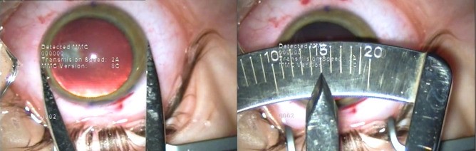
To describe the results of toric intraocular lens (IOL) implantation in three atypical cases (four eyes) with cataract and corneal astigmatism: one with bilateral keratoconus, one with pellucid marginal degeneration and one with buphthalmos due to congenital glaucoma. Three patients (four eyes) with corneal astigmatism (one with bilateral keratoconus, one with pellucid marginal degeneration and one with buphthalmos due to congenital glaucoma) underwent cataract surgery by standard phacoemulsification and the implantation of toric IOLs in the capsular bag. The presence of corneal astigmatism was identified by automated keratometry and confirmed by Scheimpflug-based corneal tomography. The toric IOL implanted in all cases was a single-piece AcrySof Toric IOL (Alcon Laboratories, Inc.). Postoperative visual acuity, the reduction in the refractive astigmatism, the spherical equivalent (SE) and the rotational stability of the toric IOL were recorded for all the patients. Visual acuity increased and the refractive astigmatism decreased in all cases. In Case 1, the right eye achieved a postoperative uncorrected visual acuity (UCVA) of 20/ 20, a decrease in the refractive astigmatism from -3 DCyl to -0.75 DCyl and a spherical equivalent (SE) of -0.25. The left eye presented with a best-corrected visual acuity (BCVA) of 20/ 20, a decrease in the refractive astigmatism from -1.50 DCyl to -1.25 DCyl and a SE of -0.25. In Case 2, the postoperative UCVA was 20/ 20, with a decrease in the refractive astigmatism from -5.5 DCyl to -1 DCyl and a SE for the right eye of 0.00 D. In Case 3, the postoperative BCVA was 20/ 20, with a decrease in the refractive astigmatism from -4.75 DCyl to -1.50 DCyl and a SE of +1.25. No misalignment of the axis of the toric IOL was observed in any patient at subsequent follow-ups. The postoperative visual acuity was satisfactory for all the patients. Toric intraocular lenses can be an effective option for implantation in patients with cataract and corneal astigmatism in atypical situations such as mild to moderate keratoconus, pellucid marginal degeneration and buphthalmos due to congenital glaucoma. Predicting the refractive outcome is difficult in atypical cases and the surgeon should have accuracy and consistency in the preoperative measurements, for achieving satisfactory postoperative results.
描述三例(四眼)白内障合并角膜散光患者(双眼圆锥角膜 1 例,角膜边缘层营养不良 1 例,先天性青光眼大泡性角膜病变 1 例)行散光型人工晶状体(toric IOL)植入的结果。三例(四眼)角膜散光患者(双眼圆锥角膜 1 例,角膜边缘层营养不良 1 例,先天性青光眼大泡性角膜病变 1 例)行白内障超声乳化摘除联合囊袋内散光型人工晶状体植入术。应用角膜自动验光仪和 Scheimpflug 角膜断层扫描仪确定角膜散光的存在。所有患者均植入 AcrySof Toric IOL(Alcon Laboratories, Inc.)。记录所有患者术后视力、屈光度、等效球镜(SE)和散光型人工晶状体旋转稳定性。所有患者术后视力均提高,屈光度降低。病例 1:右眼术后裸眼视力(UCVA)为 20/20,散光从-3.00D 减少到-0.75D,SE 为-0.25D;左眼最佳矫正视力(BCVA)为 20/20,散光从-1.50D 减少到-1.25D,SE 为-0.25D。病例 2:右眼术后 UCVA 为 20/20,散光从-5.50D 减少到-1.00D,SE 为 0.00D;左眼术后 UCVA 为 20/20,散光从-5.50D 减少到-1.00D,SE 为 0.00D。病例 3:右眼术后 BCVA 为 20/20,散光从-4.75D 减少到-1.50D,SE 为+1.25D。所有患者在随后的随访中均未发现散光型人工晶状体轴位偏位。所有患者术后视力均满意。在角膜形态异常的情况下,如轻度至中度圆锥角膜、角膜边缘层营养不良和先天性青光眼大泡性角膜病变,散光型人工晶状体植入术是治疗白内障合并角膜散光的有效选择。在这些非典型病例中,预测屈光结果较为困难,术者术前测量应准确且一致,以获得满意的术后效果。