Department of Nuclear Medicine and Clinical Molecular Imaging, University Hospital Tuebingen, Tuebingen, Germany.
Department of Diagnostic and Interventional Radiology, University Hospital Tuebingen, Tuebingen, Germany.
PLoS One. 2020 Dec 30;15(12):e0244235. doi: 10.1371/journal.pone.0244235. eCollection 2020.
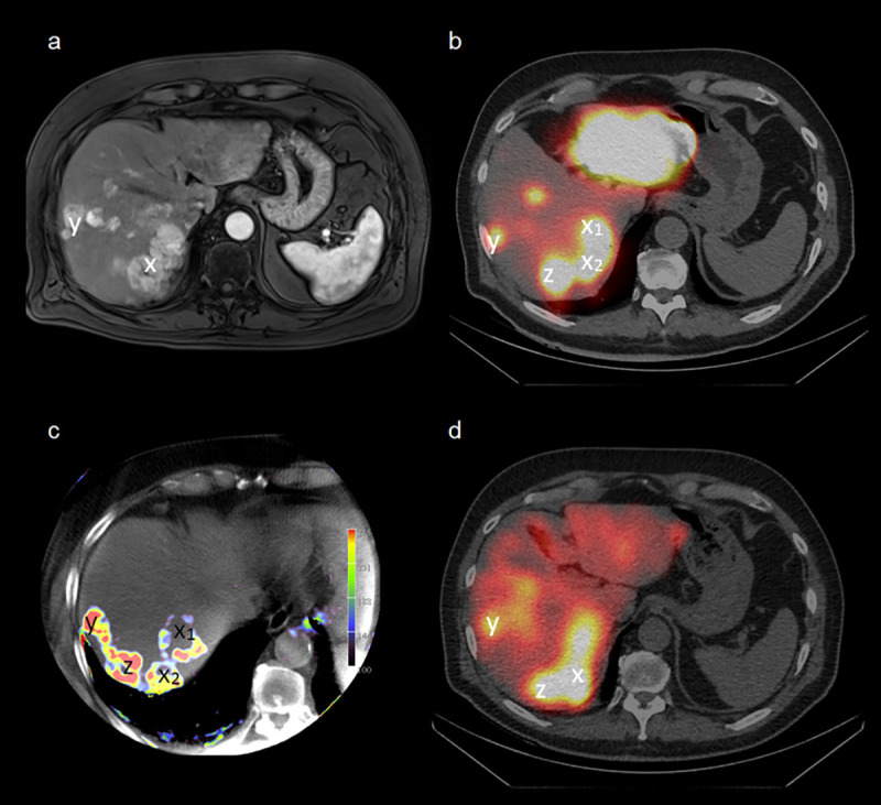
SPECT/CT with 99mTc-macroaggregated albumin (MAA) is generally used for diagnostic work-up prior to transarterial radioembolization (TARE) to exclude shunts and to provide additional information for treatment stratification and dose calculation. C-arm CT is used for determination of lobular vascular supply and assessment of parenchymal blood volume (PBV). Aim of this study was to correlate MAA-uptake and PBV-maps in hepatocellular carcinoma (HCC) and hepatic metastases of the colorectal carcinoma (CRC).
34 patients underwent a PBV C-arm CT immediately followed by 99mTc-MAA injection and a SPECT/CT acquisition after 1 h uptake. MAA-uptake and PBV-maps were visually assessed and semi-quantitatively analyzed (MAA-tumor/liver-parenchyma = MAA-TBR or PBV in ml/100ml). In case of a poor match, tumors were additionally correlated with post-TARE 90Y-Bremsstrahlung-SPECT/CT as a reference.
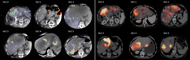
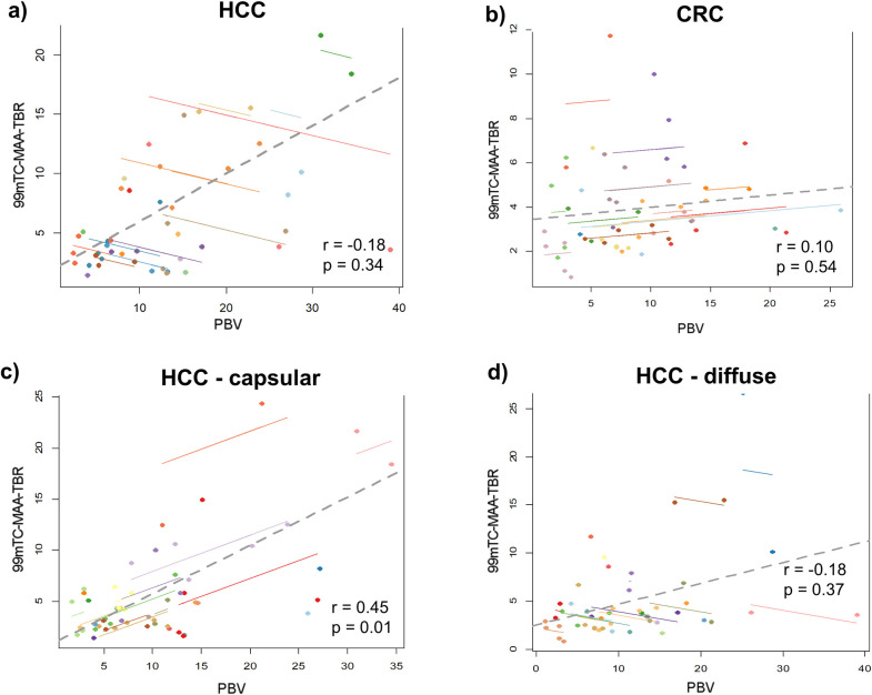
102 HCC or CRC metastases were analyzed. HCC presented with significantly higher MAA-TBR (7.6 vs. 3.9, p<0.05) compared to CRC. Tumors showed strong intra- and inter-individual dissimilarities between TBR and PBV with a weak correlations for capsular HCCs (r = 0.45, p<0.05) and no correlation for CRC. The demarcation of lesions was slightly better for both HCC and CRC in PBV-maps compared to MAA-SPECT/CT (exact match: 52%/50%; same intensity/homogeneity: 38%/39%; insufficient 10%/11%). MAA-SPECT/CT revealed a better visual correlation with post-therapeutic 90Y-Bremsstrahlung-SPECT/CT.
The acquisition of PBV can improve the detectability of small intrahepatic tumors and correlates with the MAA-Uptake in HCC. The results indicate that 99mTc-MAA-SPECT/CT remains to be the superior method for the prediction of post-therapeutic 90Y-particle distribution, especially in CRC. However, intra-procedural PBV acquisition has the potential to become an additional factor for TARE planning, in addition to improving the determination of segment and tumor blood supply, which has been demonstrated previously.
SPECT/CT 联合 99mTc-聚合白蛋白(MAA)通常用于经动脉放射栓塞(TARE)前的诊断检查,以排除分流,并为治疗分层和剂量计算提供额外信息。C 臂 CT 用于确定小叶血管供应和评估实质血容量(PBV)。本研究的目的是比较肝细胞癌(HCC)和结直肠癌(CRC)肝转移的 MAA 摄取和 PBV 图。
34 例患者行 PBV C 臂 CT 检查,随后立即注射 99mTc-MAA,1 h 后行 SPECT/CT 采集。对 MAA 摄取和 PBV 图进行视觉评估和半定量分析(MAA-肿瘤/肝实质= MAA-TBR 或 PBV 以 ml/100ml 表示)。如果匹配不佳,还将肿瘤与 TARE 后 90Y 放射性核素 Bremsstrahlung-SPECT/CT 进行额外比较。
共分析了 102 例 HCC 或 CRC 转移瘤。与 CRC 相比,HCC 的 MAA-TBR 显著更高(7.6 比 3.9,p<0.05)。肿瘤的 TBR 和 PBV 之间存在明显的个体内和个体间差异,包膜 HCC 之间相关性较弱(r=0.45,p<0.05),CRC 之间无相关性。与 MAA-SPECT/CT 相比,HCC 和 CRC 的 PBV 图在病变边界的显示上略好(完全匹配:52%/50%;相同强度/均匀性:38%/39%;不足 10%/11%)。MAA-SPECT/CT 与 TARE 后 90Y 放射性核素 Bremsstrahlung-SPECT/CT 的视觉相关性更好。
PBV 的采集可以提高小肝内肿瘤的检出率,并与 HCC 中的 MAA 摄取相关。结果表明,99mTc-MAA-SPECT/CT 仍然是预测 TARE 后 90Y 粒子分布的较好方法,尤其是在 CRC 中。然而,与之前已经证明的确定节段和肿瘤血供的作用相比,术中 PBV 采集有可能成为 TARE 计划的附加因素。