Yanaka Kenichi, Konishi Akihide, Shinke Toshiro, Kozuki Amane, Kawamori Hiroyuki, Tsukiyama Yoshiro, Iida Osamu, Kadotani Makoto, Omori Takashi, Hirata Ken-Ichi
Division of Cardiovascular Medicine, Department of Internal Medicine, Kobe University Graduate School of Medicine.
Clinical & Translational Research Center, Kobe University Hospital.
Ann Vasc Dis. 2020 Sep 25;13(3):291-299. doi: 10.3400/avd.oa.20-00077.
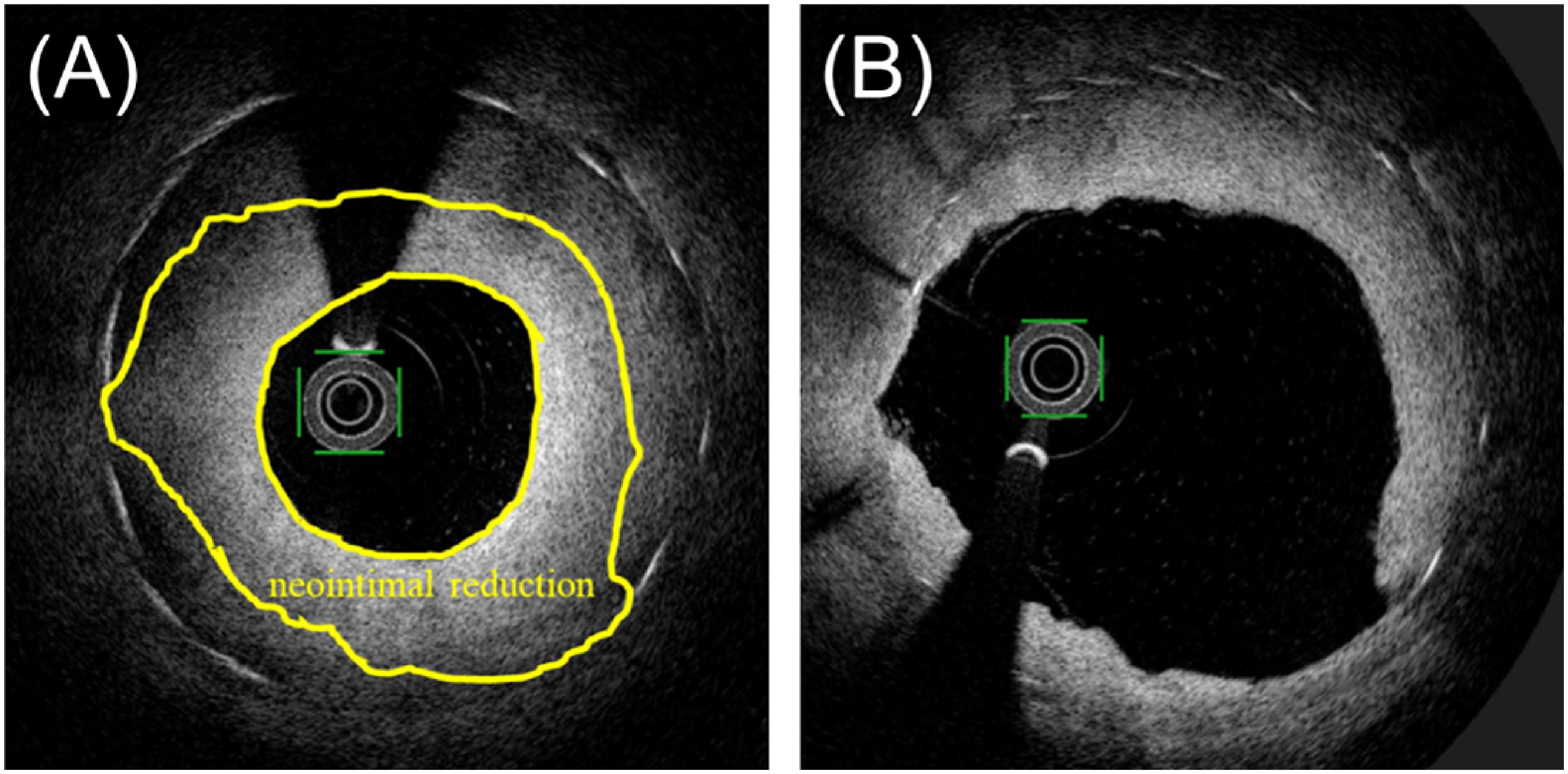
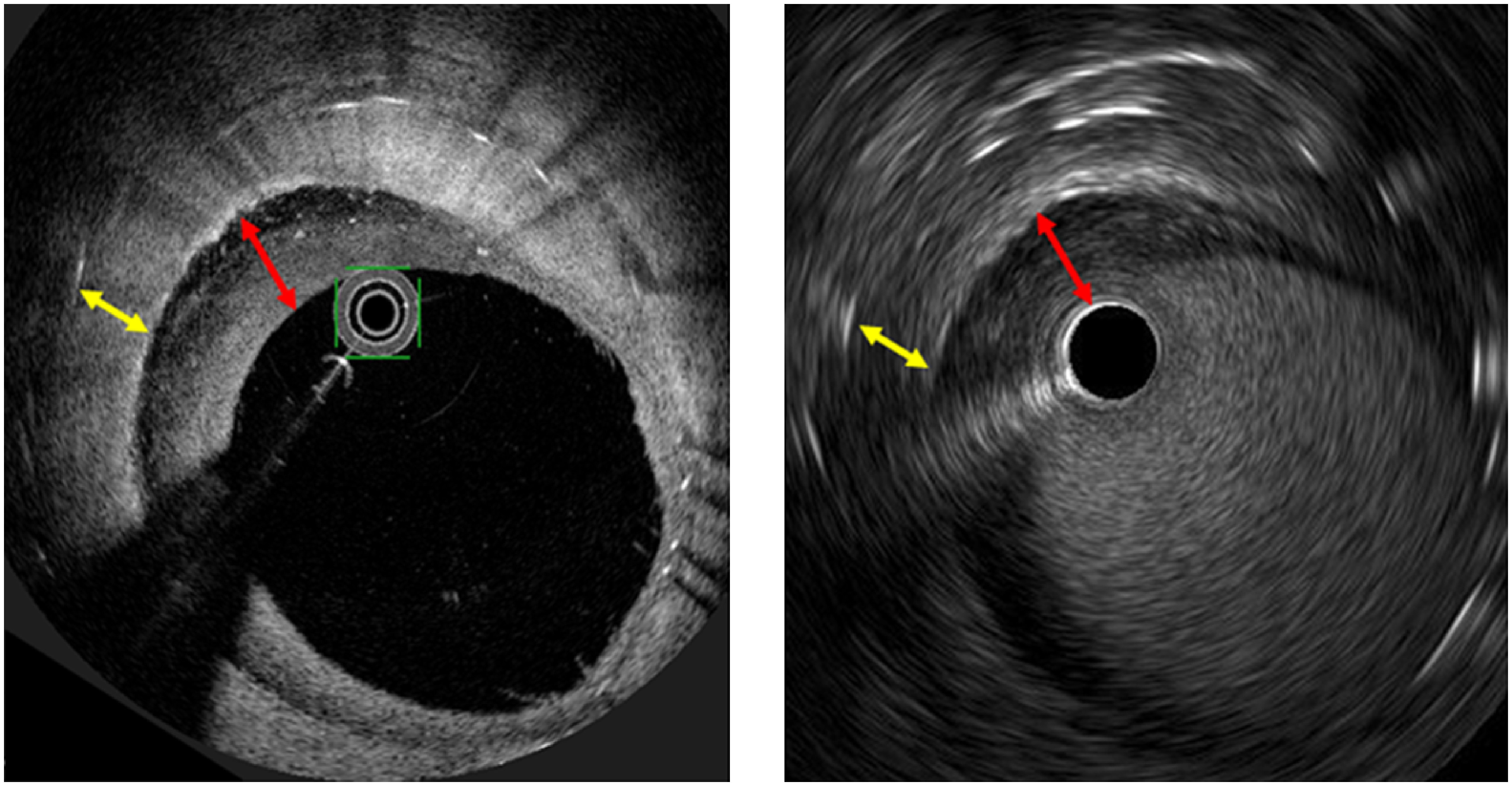
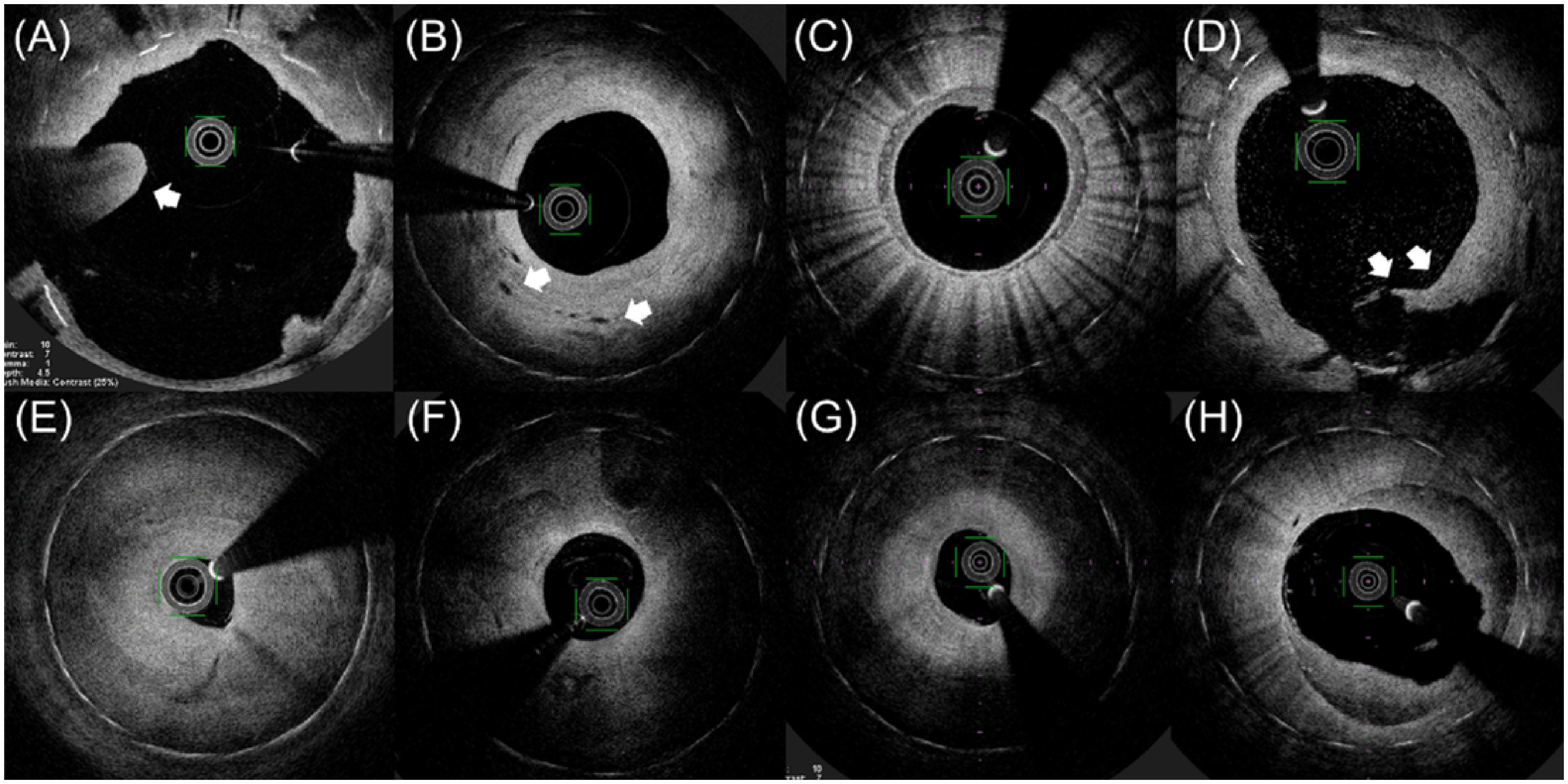
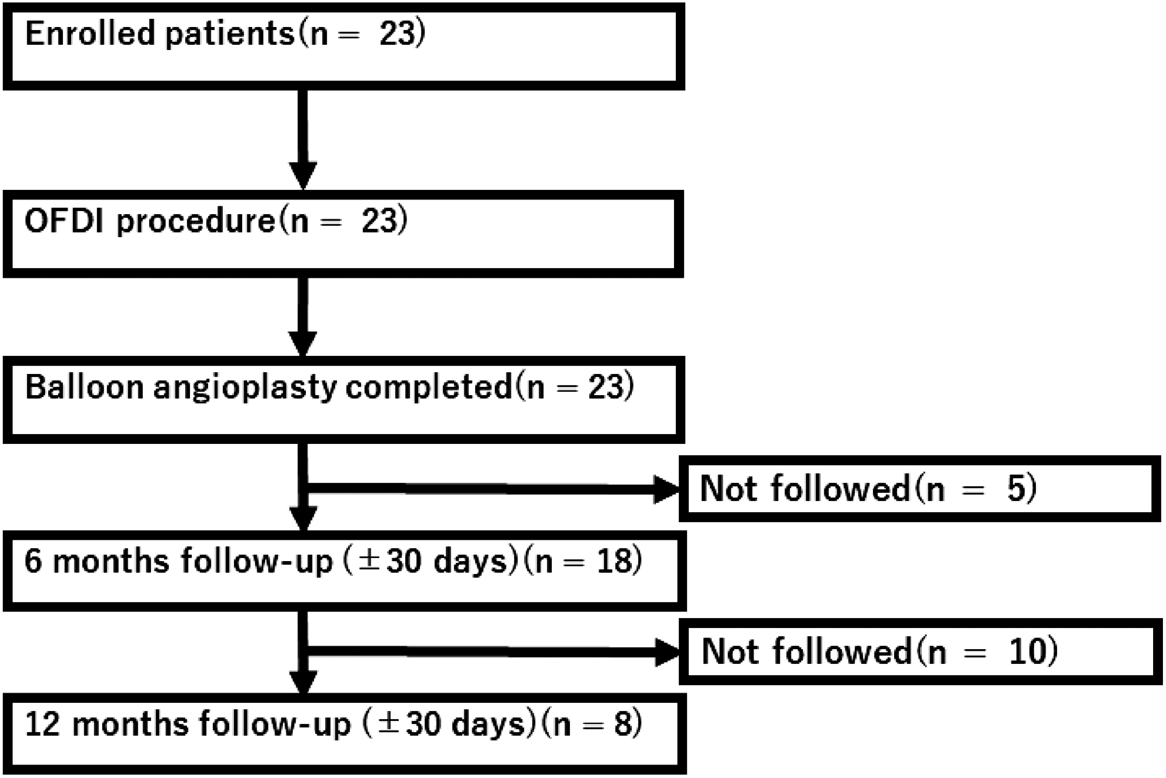
: Balloon angioplasty for in-stent restenosis (ISR) in the superficial femoral artery (SFA) has a high recurrent restenosis rate; however, its mechanism has not been fully and precisely evaluated using high-resolution intravascular imaging. Thus, we aimed to evaluate the relationship between vascular features obtained by optical frequency domain imaging (OFDI) and recurrent restenosis at 6 months. : This was a prospective multicenter single-arm study. OFDI was performed before and after balloon angioplasty, and vascular features were assessed. A multi-layered ISR pattern detected by OFDI was defined as several signal-poor appearances with a high-signal band adjacent to the luminal surface. The primary outcome was defined as recurrent restenosis 6 months after balloon angioplasty. : Given that this study was terminated early, only 18 patients completed the 6-month follow-up; of these, 8 developed restenosis. Recurrent restenosis at 6 months tended to be related to a multi-layered ISR pattern (odds ratio (OR), 6.67; 95% confidence interval (CI), 0.81-54.96; p=0.078) and the minimum lumen area (MLA) after balloon angioplasty (OR, 0.71; 95%CI, 0.48-1.04; p=0.077). : A multi-layered ISR pattern and MLA after balloon angioplasty detected by OFDI might be risk factors for recurrent ISR in the SFA.
股浅动脉(SFA)支架内再狭窄(ISR)的球囊血管成形术复发性再狭窄率较高;然而,其机制尚未通过高分辨率血管内成像进行全面而精确的评估。因此,我们旨在评估光学频域成像(OFDI)获得的血管特征与6个月时复发性再狭窄之间的关系。
这是一项前瞻性多中心单臂研究。在球囊血管成形术前后进行OFDI,并评估血管特征。OFDI检测到的多层ISR模式定义为与管腔表面相邻的几个信号较弱外观以及一个高信号带。主要结局定义为球囊血管成形术6个月后的复发性再狭窄。
鉴于本研究提前终止,只有18例患者完成了6个月的随访;其中8例发生了再狭窄。6个月时的复发性再狭窄倾向于与多层ISR模式(优势比(OR),6.67;95%置信区间(CI),0.81 - 54.96;p = 0.078)以及球囊血管成形术后的最小管腔面积(MLA)(OR,0.71;95%CI,0.48 - 1.04;p = 0.077)相关。
OFDI检测到的球囊血管成形术后的多层ISR模式和MLA可能是SFA复发性ISR的危险因素。