Sitta Juliana, Howard Candace M
Department of Radiology, University of Mississippi Medical Center, 2500 North State Street Jackson, Mississippi 39216, USA.
Radiol Case Rep. 2020 Dec 22;16(3):538-542. doi: 10.1016/j.radcr.2020.12.028. eCollection 2021 Mar.
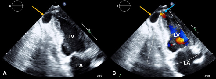
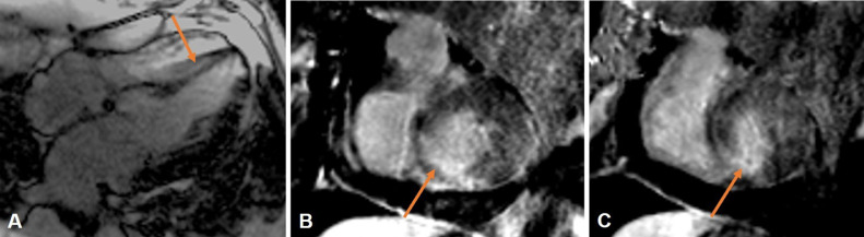
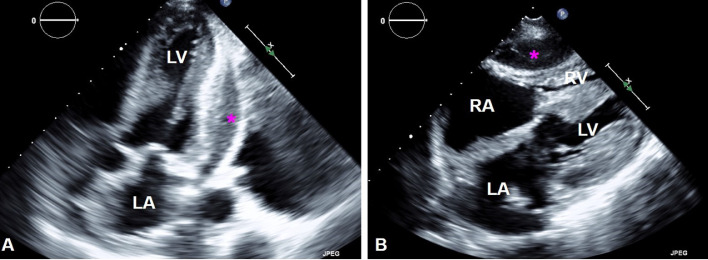
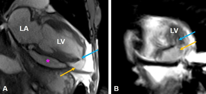
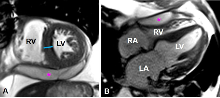
A left ventricle pseudoaneurysm (LV PSA) is defined as a free wall rupture of the left ventricle contained by the adjacent pericardial tissue. This rare complication is most commonly encountered following myocardial infarction, trauma, or infection. Surgery is typically warranted to avoid progression to spontaneous rupture, which may potentially lead to cardiac tamponade and death. Cardiac magnetic resonance imaging is the modality of choice to characterize left ventricle morphology and function. Accurate distinction between a pseudoaneurysm and a true aneurysm is crucial, since management and prognosis are significantly different between these 2 entities. We present a case of a 63-year-old male heart transplant recipient, admitted for suspicion of acute cellular rejection, with an unexpected finding of a LV PSA.
左心室假性动脉瘤(LV PSA)定义为左心室游离壁破裂,由相邻心包组织包裹。这种罕见的并发症最常见于心肌梗死、创伤或感染后。通常需要进行手术以避免进展为自发性破裂,后者可能导致心脏压塞和死亡。心脏磁共振成像是描述左心室形态和功能的首选检查方法。准确区分假性动脉瘤和真性动脉瘤至关重要,因为这两种情况的治疗和预后有显著差异。我们报告一例63岁男性心脏移植受者,因疑似急性细胞排斥反应入院,意外发现左心室假性动脉瘤。