Department of Nuclear Medicine, Peking Union Medical College Hospital, Chinese Academy of Medical Science & Peking Union Medical College, Beijing Key Laboratory of Molecular Targeted Diagnosis and Therapy in Nuclear Medicine, Beijing, China.
Radiochemistry and Radiation Chemistry Key Laboratory of Fundamental Science, Beijing National Laboratory for Molecular Sciences, College of Chemistry and Molecular Engineering, Peking University, Beijing, 100871, China.
Theranostics. 2021 Jan 1;11(1):304-315. doi: 10.7150/thno.45540. eCollection 2021.
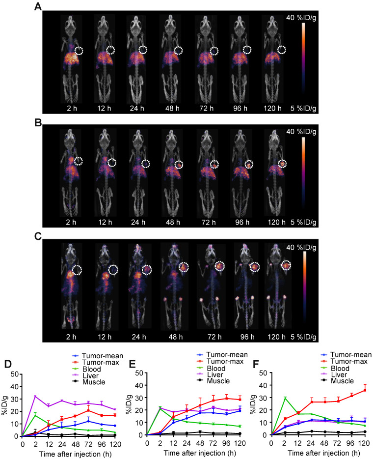
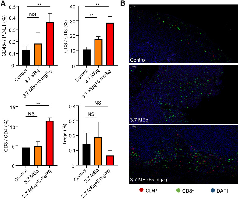
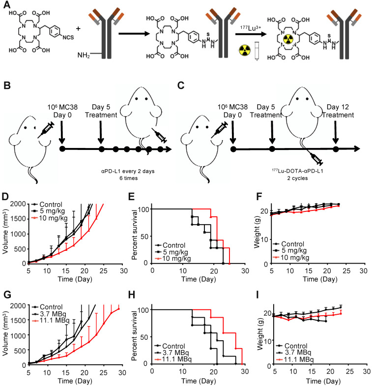
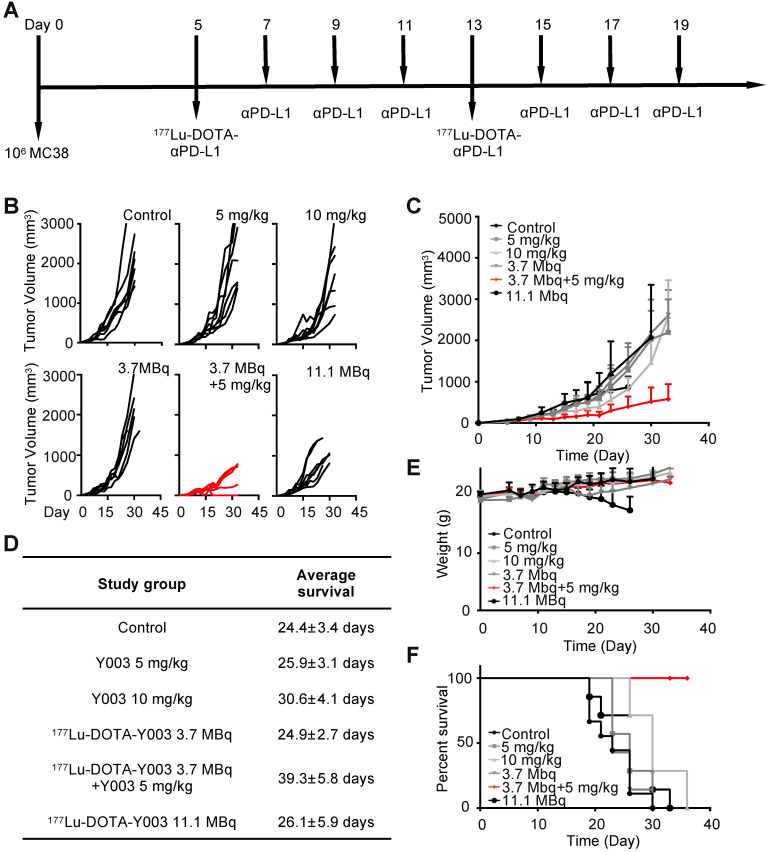
The low response rate of immunotherapy, such as anti-PD-L1/PD-1 and anti-CTLA4, has limited its application to a wider population of cancer patients. One widely accepted view is that inflammation within the tumor microenvironment is low or ineffective for inducing the sufficient infiltration and/or activation of lymphocytes. Here, a highly tumor-selective anti-PD-L1 (αPD-L1) antibody was developed through PET imaging screening, and it was radiolabeled with Lu-177 for PD-L1-targeted radioimmunotherapy (RIT) and radiation-synergized immunotherapy. A series of αPD-L1 antibodies were radiolabeled with zirconium-89 for PET imaging to screen the most suitable antibodies for RIT. Mice were divided into an immunotherapy group, a RIT group and a radiation-synergized immunotherapy group to evaluate the therapeutic effect. Alterations in the tumor microenvironment after treatment were assessed using flow cytometry and immunofluorescence microscopy. Radiation-synergistic RIT can achieve a significantly better therapeutic effect than immunotherapy or RIT alone. The dosages of the radiopharmaceuticals and αPD-L1 antibodies were reduced, the infiltration of CD4 and CD8 T cells in the tumor microenvironment was increased, and no side effects were observed. This radiation-synergistic RIT strategy successfully showed a strong synergistic effect with αPD-L1 checkpoint blockade therapy, at least in the mouse model. PET imaging of Zr-labeled antibodies is an effective method for antibody screening. RIT with a Lu-labeled αPD-L1 antibody could successfully upregulate antitumor immunity in the tumor microenvironment and turn "cold" tumors "hot" for immunotherapy.
免疫疗法(如抗 PD-L1/PD-1 和抗 CTLA4)的低应答率限制了其在更广泛的癌症患者群体中的应用。一种被广泛接受的观点是,肿瘤微环境中的炎症水平低或无效,无法诱导足够的淋巴细胞浸润和/或激活。在这里,通过 PET 成像筛选开发了一种高度肿瘤选择性的抗 PD-L1(αPD-L1)抗体,并将其用镥-177 标记用于 PD-L1 靶向放射免疫治疗(RIT)和放射增敏免疫治疗。一系列αPD-L1 抗体用锆-89 标记用于 PET 成像,以筛选最适合 RIT 的抗体。将小鼠分为免疫治疗组、RIT 组和放射增敏免疫治疗组,以评估治疗效果。使用流式细胞术和免疫荧光显微镜评估治疗后肿瘤微环境的变化。放射增敏 RIT 可实现明显优于免疫治疗或单独 RIT 的治疗效果。减少了放射性药物和αPD-L1 抗体的剂量,增加了肿瘤微环境中 CD4 和 CD8 T 细胞的浸润,并且没有观察到副作用。这种放射增敏 RIT 策略在小鼠模型中至少成功地显示出与αPD-L1 检查点阻断治疗的强烈协同作用。Zr 标记抗体的 PET 成像是一种有效的抗体筛选方法。用 Lu 标记的αPD-L1 抗体进行 RIT 可以成功上调肿瘤微环境中的抗肿瘤免疫,使“冷”肿瘤对免疫治疗“热”。