• 文献检索
  • 文档翻译
  • 深度研究
  • 学术资讯
  • Suppr Zotero 插件Zotero 插件
  • 邀请有礼
  • 套餐&价格
  • 历史记录
应用&插件
Suppr Zotero 插件Zotero 插件浏览器插件Mac 客户端Windows 客户端微信小程序
定价
高级版会员购买积分包购买API积分包
服务
文献检索文档翻译深度研究API 文档MCP 服务
关于我们
关于 Suppr公司介绍联系我们用户协议隐私条款
关注我们

Suppr 超能文献

核心技术专利:CN118964589B侵权必究
粤ICP备2023148730 号-1Suppr @ 2026

文献检索

告别复杂PubMed语法,用中文像聊天一样搜索,搜遍4000万医学文献。AI智能推荐,让科研检索更轻松。

立即免费搜索

文件翻译

保留排版,准确专业,支持PDF/Word/PPT等文件格式,支持 12+语言互译。

免费翻译文档

深度研究

AI帮你快速写综述,25分钟生成高质量综述,智能提取关键信息,辅助科研写作。

立即免费体验

小儿大气道疾病:图文综述

Large airway diseases in pediatrics: a pictorial essay.

作者信息

Prountzos Spyridon, Papakonstantinou Olympia, Bizimi Vasiliki, Velonakis Georgios, Mazioti Argyro, Douros Konstantinos, Alexopoulou Efthymia

机构信息

2nd Department of Radiology, National and Kapodistrian University of Athens, "Attikon" University Hospital of Athens, Athens, Greece.

Allergology and Pulmonology Unit, 3rd Pediatric Department, Attikon Hospital, National and Kapodistrian University of Athens, Athens, Greece.

出版信息

Acta Radiol Open. 2020 Dec 16;9(12):2058460120972694. doi: 10.1177/2058460120972694. eCollection 2020 Dec.

DOI:10.1177/2058460120972694
PMID:33403124
原文链接:https://pmc.ncbi.nlm.nih.gov/articles/PMC7747119/
Abstract

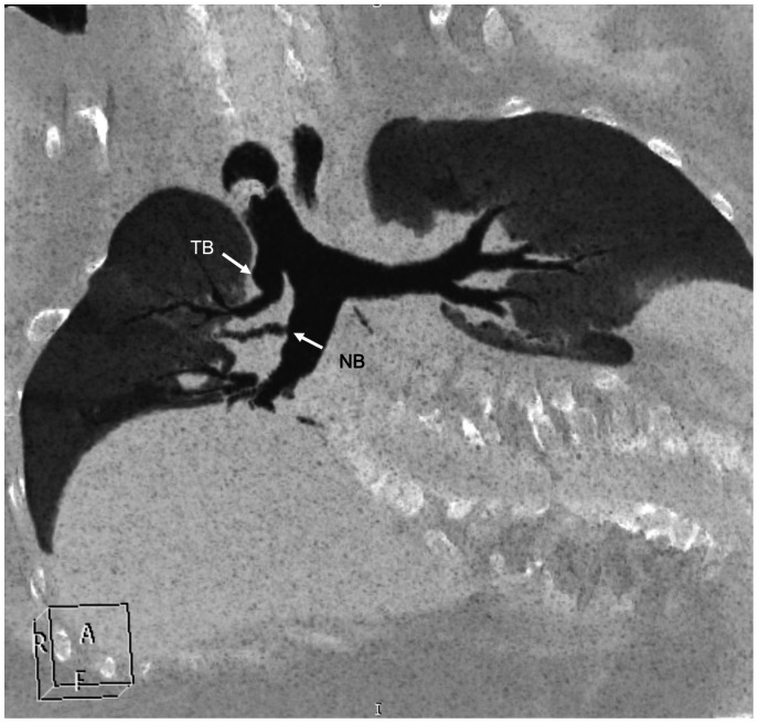
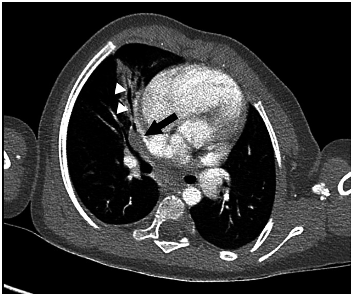
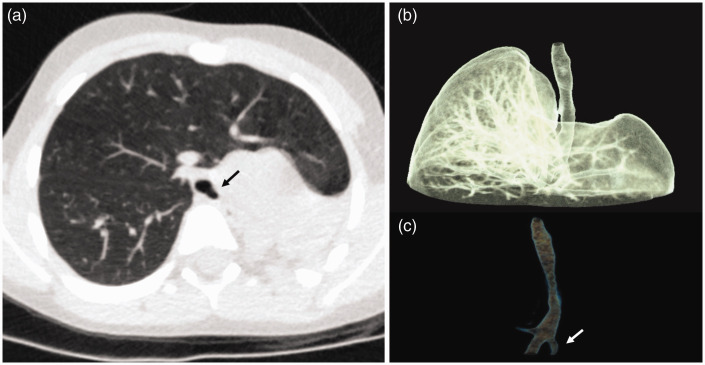
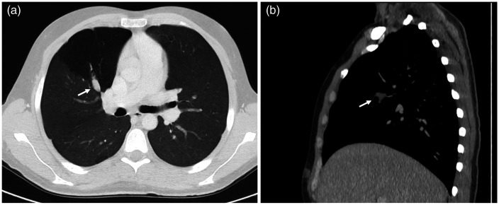
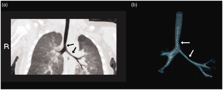
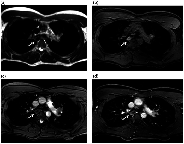
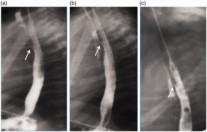
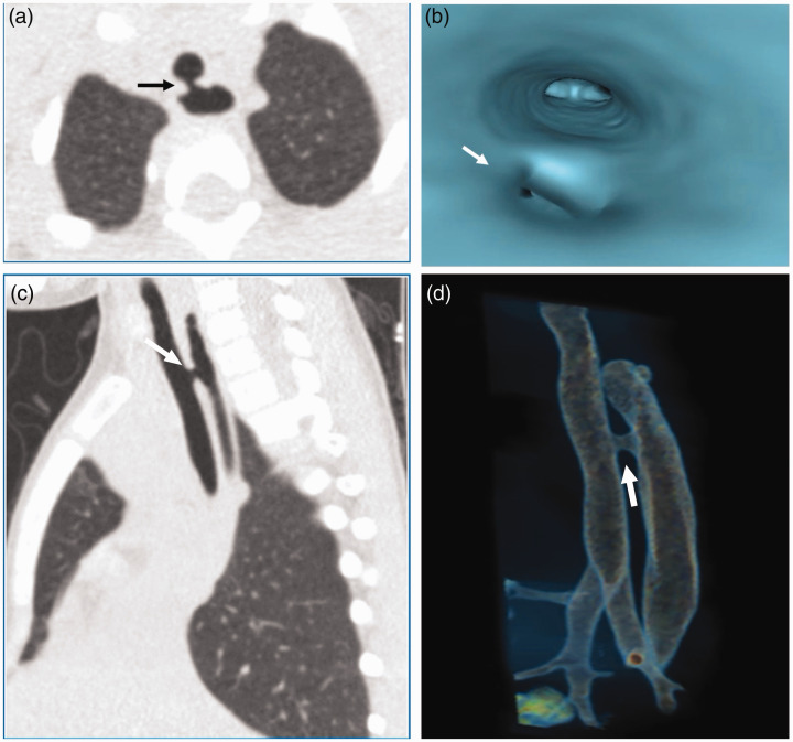
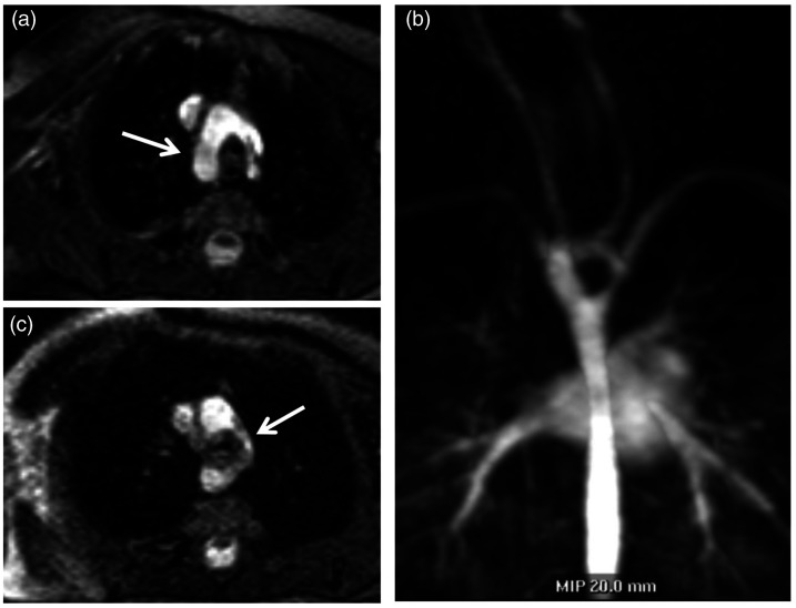
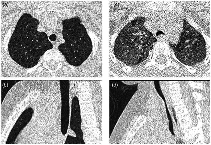
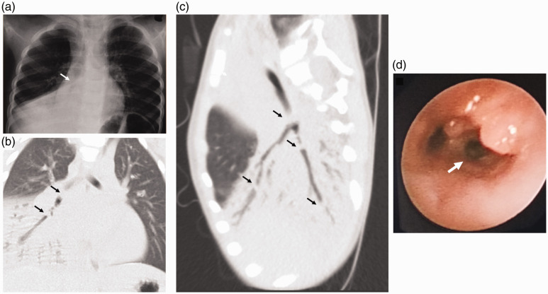
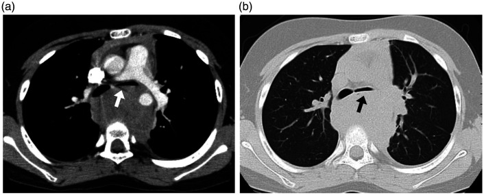
"Large airway diseases" is being used as an all-encompassing phrase to describe a broad spectrum of pathological entities, which involves the trachea, main, lobar, and segmental bronchi of up to 3 mm diameter. Imaging modalities such as radiography, computed tomography, and magnetic resonance imaging contribute to the identification and diagnosis of each entity. Knowledge of clinical information, normal cross-sectional anatomy, and imaging characteristics of large airway diseases is necessary for appropriate radiologic evaluation. This review provides information about congenital and acquired diseases of the large airways in the pediatric population.

摘要

“大气道疾病”被用作一个涵盖性术语,用于描述一系列广泛的病理实体,这些实体涉及气管、直径达3毫米的主支气管、叶支气管和段支气管。诸如X线摄影、计算机断层扫描和磁共振成像等成像方式有助于识别和诊断每个实体。了解临床信息、正常横断面解剖结构以及大气道疾病的影像学特征对于进行适当的放射学评估是必要的。本综述提供了有关儿科人群大气道先天性和后天性疾病的信息。

https://cdn.ncbi.nlm.nih.gov/pmc/blobs/8077/7747119/7a77c46bd3cb/10.1177_2058460120972694-fig14.jpg
https://cdn.ncbi.nlm.nih.gov/pmc/blobs/8077/7747119/bb72cc0bdcd1/10.1177_2058460120972694-fig1.jpg
https://cdn.ncbi.nlm.nih.gov/pmc/blobs/8077/7747119/ad77dfea7527/10.1177_2058460120972694-fig2.jpg
https://cdn.ncbi.nlm.nih.gov/pmc/blobs/8077/7747119/68ec70c12f60/10.1177_2058460120972694-fig3.jpg
https://cdn.ncbi.nlm.nih.gov/pmc/blobs/8077/7747119/da8f38efebf1/10.1177_2058460120972694-fig4.jpg
https://cdn.ncbi.nlm.nih.gov/pmc/blobs/8077/7747119/be8047e5244e/10.1177_2058460120972694-fig5.jpg
https://cdn.ncbi.nlm.nih.gov/pmc/blobs/8077/7747119/2800ff7f4922/10.1177_2058460120972694-fig6.jpg
https://cdn.ncbi.nlm.nih.gov/pmc/blobs/8077/7747119/07837008bbbd/10.1177_2058460120972694-fig7.jpg
https://cdn.ncbi.nlm.nih.gov/pmc/blobs/8077/7747119/766e27f509d5/10.1177_2058460120972694-fig8.jpg
https://cdn.ncbi.nlm.nih.gov/pmc/blobs/8077/7747119/7478870b1630/10.1177_2058460120972694-fig9.jpg
https://cdn.ncbi.nlm.nih.gov/pmc/blobs/8077/7747119/5b7dd62963f1/10.1177_2058460120972694-fig10.jpg
https://cdn.ncbi.nlm.nih.gov/pmc/blobs/8077/7747119/50c467b45e3b/10.1177_2058460120972694-fig11.jpg
https://cdn.ncbi.nlm.nih.gov/pmc/blobs/8077/7747119/c69355e13618/10.1177_2058460120972694-fig12.jpg
https://cdn.ncbi.nlm.nih.gov/pmc/blobs/8077/7747119/e28b93c1a696/10.1177_2058460120972694-fig13.jpg
https://cdn.ncbi.nlm.nih.gov/pmc/blobs/8077/7747119/7a77c46bd3cb/10.1177_2058460120972694-fig14.jpg

相似文献

1
Large airway diseases in pediatrics: a pictorial essay.小儿大气道疾病:图文综述
Acta Radiol Open. 2020 Dec 16;9(12):2058460120972694. doi: 10.1177/2058460120972694. eCollection 2020 Dec.
2
Current and future approaches to large airways imaging in adults and children.成人和儿童大气道成像的当前及未来方法
Clin Radiol. 2017 May;72(5):356-374. doi: 10.1016/j.crad.2017.01.012. Epub 2017 Mar 1.
3
Multi-detector computed tomography imaging of large airway pathology: A pictorial review.大气道病变的多排螺旋计算机断层扫描成像:图谱综述。
World J Radiol. 2015 Dec 28;7(12):459-74. doi: 10.4329/wjr.v7.i12.459.
4
Computed tomography findings of tracheobronchial system diseases: a pictorial essay.气管支气管系统疾病的计算机断层扫描表现:图文综述
Jpn J Radiol. 2015 Feb;33(2):51-8. doi: 10.1007/s11604-014-0380-3. Epub 2014 Dec 11.
5
Pulmonary Valve Anatomy and Abnormalities: A Pictorial Essay of Radiography, Computed Tomography (CT), and Magnetic Resonance Imaging (MRI).肺动脉瓣解剖与异常:X线摄影、计算机断层扫描(CT)及磁共振成像(MRI)的图文综述
J Thorac Imaging. 2016 Jan;31(1):W4-12. doi: 10.1097/RTI.0000000000000182.
6
Multimodality imaging of pediatric airways disease: indication and technique.儿童气道疾病的多模态成像:适应证与技术
Radiol Med. 2017 Jun;122(6):419-429. doi: 10.1007/s11547-017-0737-7. Epub 2017 Feb 21.
7
Multidetector computed tomography imaging of congenital anomalies of major airways: A pictorial essay.多排螺旋计算机断层扫描对大气道先天性异常的成像:图文综述。
World J Radiol. 2011 Dec 28;3(12):289-97. doi: 10.4329/wjr.v3.i12.289.
8
Grading airway stenosis down to the segmental level using virtual bronchoscopy.使用虚拟支气管镜检查将气道狭窄分级至节段水平。
Chest. 2004 Feb;125(2):704-11. doi: 10.1378/chest.125.2.704.
9
[Specific characteristics of chest X‑ray in childhood : Basics for radiologists].[儿童胸部X线的特殊特征:放射科医生的基础]
Radiologe. 2018 Apr;58(4):359-376. doi: 10.1007/s00117-018-0374-8.
10
CT Findings of Central Airway Lesions Causing Airway Stenosis-Visualization and Quantification: A Pictorial Essay.导致气道狭窄的中央气道病变的CT表现——可视化与量化:一篇图文并茂的文章
Taehan Yongsang Uihakhoe Chi. 2021 Nov;82(6):1441-1476. doi: 10.3348/jksr.2020.0212. Epub 2021 Sep 16.

引用本文的文献

1
Forty days old infant with Pig Bronchus, presenting with recurrent pneumonia: A Case Report.40日龄婴儿患猪支气管,反复发生肺炎:病例报告
Pediatric Health Med Ther. 2023 Oct 30;14:379-383. doi: 10.2147/PHMT.S429852. eCollection 2023.

本文引用的文献

1
Avoiding unnecessary bronchoscopy in children with suspected foreign body aspiration using computed tomography.使用计算机断层扫描避免怀疑异物吸入的儿童进行不必要的支气管镜检查。
J Pediatr Surg. 2020 Jan;55(1):176-181. doi: 10.1016/j.jpedsurg.2019.09.045. Epub 2019 Oct 25.
2
ERS statement on tracheomalacia and bronchomalacia in children.ERS 关于儿童气管软化和支气管软化的声明。
Eur Respir J. 2019 Sep 28;54(3). doi: 10.1183/13993003.00382-2019. Print 2019 Sep.
3
Solitary round pulmonary lesions in the pediatric population: a pictorial review.
儿科人群中的孤立性圆形肺病变:影像学综述
Acta Radiol Open. 2019 May 31;8(5):2058460119851998. doi: 10.1177/2058460119851998. eCollection 2019 May.
4
Pediatric Mediastinal Masses:: Role of MR Imaging as a Problem-Solving Tool.小儿纵隔肿块:磁共振成像作为问题解决工具的作用
Magn Reson Imaging Clin N Am. 2019 May;27(2):227-242. doi: 10.1016/j.mric.2019.01.003.
5
Pediatric Malignant Mediastinal Masses.小儿恶性纵隔肿块
J Coll Physicians Surg Pak. 2019 Mar;29(3):258-262. doi: 10.29271/jcpsp.2019.03.258.
6
Four-Dimensional Thoracic CT in Free-Breathing Children.自由呼吸状态下儿童的四维胸部 CT
Korean J Radiol. 2019 Jan;20(1):50-57. doi: 10.3348/kjr.2018.0325. Epub 2018 Dec 27.
7
Helical multi-detector CT scan as a tool for diagnosing tracheomalacia in children.螺旋多排 CT 扫描在儿童气管软化症诊断中的应用。
Pediatr Pulmonol. 2019 Jan;54(1):47-52. doi: 10.1002/ppul.24188. Epub 2018 Nov 28.
8
Current and future approaches to large airways imaging in adults and children.成人和儿童大气道成像的当前及未来方法
Clin Radiol. 2017 May;72(5):356-374. doi: 10.1016/j.crad.2017.01.012. Epub 2017 Mar 1.
9
Multimodality imaging of pediatric airways disease: indication and technique.儿童气道疾病的多模态成像:适应证与技术
Radiol Med. 2017 Jun;122(6):419-429. doi: 10.1007/s11547-017-0737-7. Epub 2017 Feb 21.
10
Multidetector Computed Tomography for Congenital Anomalies of the Aortic Arch: Vascular Rings.多层螺旋计算机断层扫描用于主动脉弓先天性异常:血管环
Rev Esp Cardiol (Engl Ed). 2016 Jul;69(7):681-93. doi: 10.1016/j.rec.2016.03.025. Epub 2016 Jun 13.