Department of Radiology the Third Hospital, Hebei Medical University, 139 Ziqiang Road, 050051, Shijiazhuang, Hebei Province, China.
BMC Musculoskelet Disord. 2021 Jan 6;22(1):27. doi: 10.1186/s12891-020-03882-2.
To investigate the imaging features of hemangiomas in long tabular bones for better diagnosis.
Twenty-four patients with long bone hemangiomas confirmed by pathology were enrolled. Nineteen patients had plain radiography, fourteen patients had computed tomography (CT) and eleven had magnetic resonance imaging (MRI). The hemangioma was divided into medullary [13], periosteal [6] and intracortical type [5].
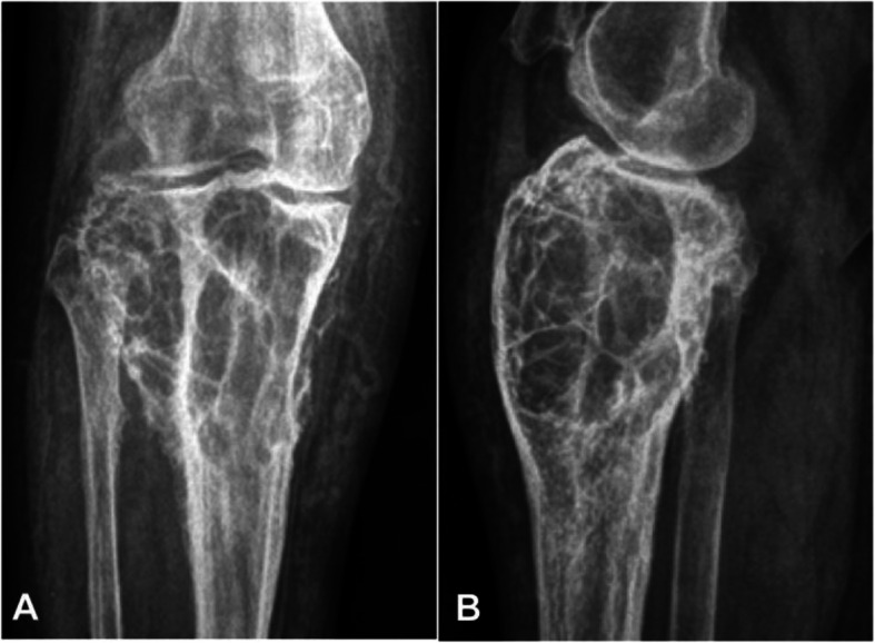
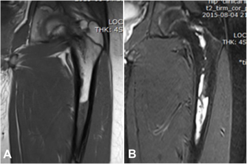
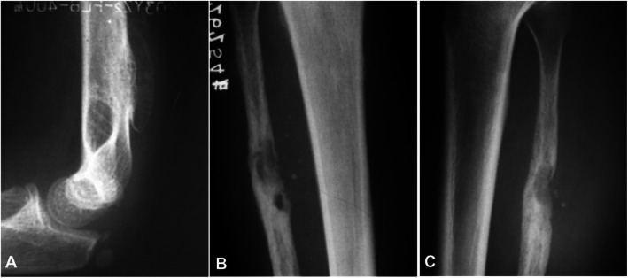
Among 19 patients with plain radiography, eleven patients were medullary, three periosteal, and five intracortical. In the medullary type, the lesion was primarily osteolytic, including five cases with irregular and unclear rims and one lesion having osteosclerotic and unclear rims. In three patients with the periosteal type, the lesion had clear rims with involvement of the cortical bone in the form of bone defect, including two cases with local thickened bone periosteum and one case having expansile periosteum. Five intracortical hemangiomas had intracortical osteolytic lesions with clear margins. Among 14 patients with CT imaging, 8 cases were medullary, three periosteal, and three intracortical. Among 8 medullary hemangiomas, one had ground glass opacity, and seven had osteolytic, expansile lesions like soft tissue density with no calcification. In three periosteal cases, the lesion was osteolytic with thickened periosteum and narrowed medullary cavity. In three intracortical hemangiomas, the lesion was of even soft tissue density with no calcification. Among 11 patients with MRI imaging, seven were medullary, two periosteal, and two intracortical. Among 7 medullary lesions, six were of hypointense signal on T1WI and hyperintensesignal on T2 WI. In two periosteal cases, the periosteum was thickened, with one case being of equal signal, and the other having no signal. Two intracortical hemangiomas were both of slightly low signal on T1WI but hyperintense signal on T2WI.
The long bone hemangiomas had characteristic cystic honeycomb-like presentations in plain radiograph. CT and MRI imagings are helpful for diagnosis of hemangiomas in long bone.
为了更好地诊断长管状骨血管瘤的影像学特征。
纳入 24 例经病理证实的长管状骨血管瘤患者。19 例行平片检查,14 例行 CT 检查,11 例行 MRI 检查。将血管瘤分为髓内型[13]、骨膜型[6]和皮质内型[5]。
平片检查 19 例中,髓内型 11 例,骨膜型 3 例,皮质内型 5 例。髓内型病变主要为溶骨性,其中 5 例边缘不规则、不清晰,1 例呈硬化性、边缘不清晰。骨膜型 3 例,边缘清晰,骨皮质受累呈骨缺损,其中 2 例骨膜增厚,1 例骨膜膨胀。5 例皮质内血管瘤表现为皮质内溶骨性病变,边缘清晰。CT 成像 14 例中,髓内型 8 例,骨膜型 3 例,皮质内型 3 例。8 例髓内血管瘤中,1 例呈磨玻璃样密度,7 例呈溶骨性、膨胀性软组织密度,无钙化。3 例骨膜型病变呈溶骨性,骨膜增厚,骨髓腔变窄。3 例皮质内血管瘤呈均匀软组织密度,无钙化。MRI 成像 11 例中,髓内型 7 例,骨膜型 2 例,皮质内型 2 例。7 例髓内病变 T1WI 呈低信号,T2WI 呈高信号。2 例骨膜型病变骨膜增厚,1 例等信号,另 1 例无信号。2 例皮质内血管瘤 T1WI 呈稍低信号,T2WI 呈高信号。
长管状骨血管瘤在平片上呈特征性的囊状蜂窝状表现。CT 和 MRI 成像有助于长骨血管瘤的诊断。