Department of Dermatology, Venereology & Allergology, Medical University of Innsbruck, Innsbruck, Austria.
Department of Immunology, St. Jude Children's Research Hospital, Memphis, Tennessee, USA.
J Immunother Cancer. 2021 Jan;9(1). doi: 10.1136/jitc-2020-000832.
Immunotherapy with checkpoint inhibitors has shown impressive results in patients with melanoma, but still many do not benefit from this line of treatment. A lack of tumor-infiltrating T cells is a common reason for therapy failure but also a loss of intratumoral dendritic cells (DCs) has been described.
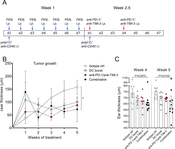
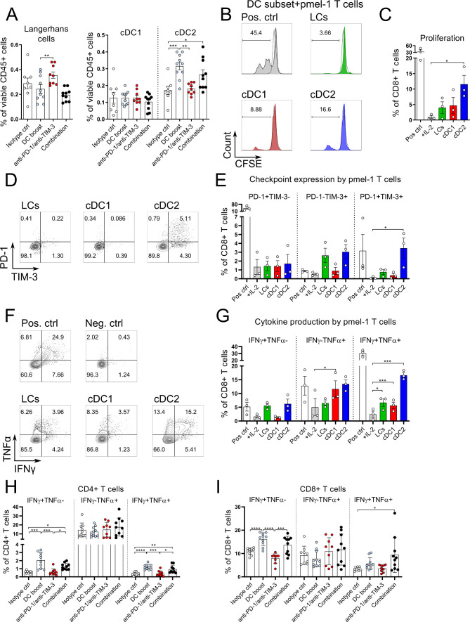
We used the transgenic tg(Grm1)EPv melanoma mouse strain that develops spontaneous, slow-growing tumors to perform immunological analysis during tumor progression. With flow cytometry, the frequencies of DCs and T cells at different tumor stages and the expression of the inhibitory molecules programmed cell death protein-1 (PD-1) and T-cell immunoglobulin and mucin-domain containing-3 (TIM-3) on T cells were analyzed. This was complemented with RNA-sequencing (RNA-seq) and real-time quantitative PCR (RT-qPCR) analysis to investigate the immune status of the tumors. To boost DC numbers and function, we administered Fms-related tyrosine 3 ligand (Flt3L) plus an adjuvant mix of polyI:C and anti-CD40. To enhance T cell function, we tested several checkpoint blockade antibodies. Immunological alterations were characterized in tumor and tumor-draining lymph nodes (LNs) by flow cytometry, CyTOF, microarray and RT-qPCR to understand how immune cells can control tumor growth. The specific role of migratory skin DCs was investigated by coculture of sorted DC subsets with melanoma-specific CD8+ T cells.
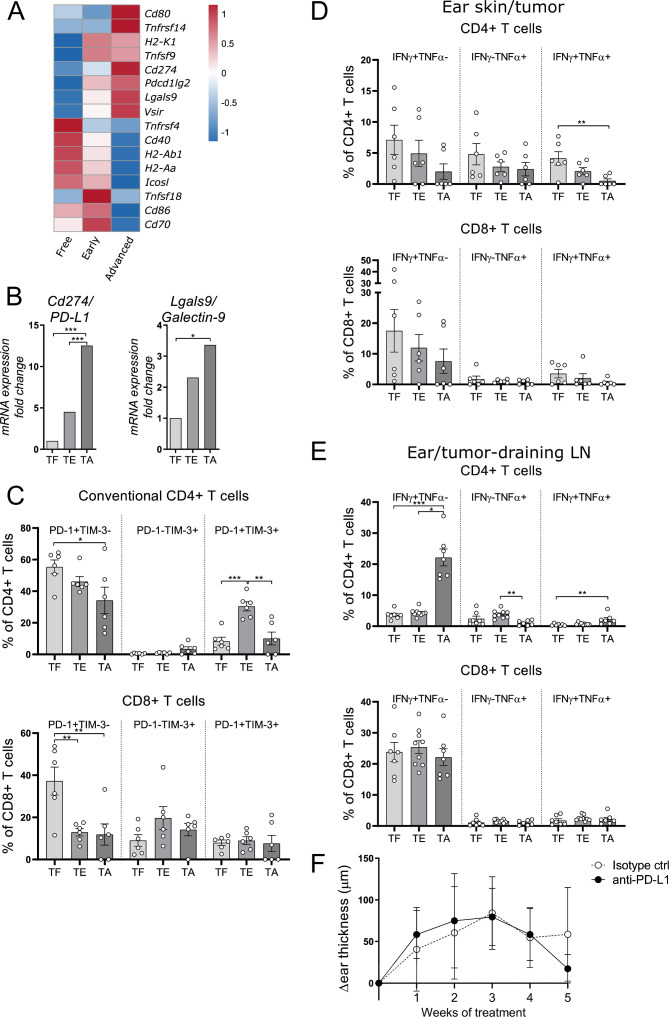
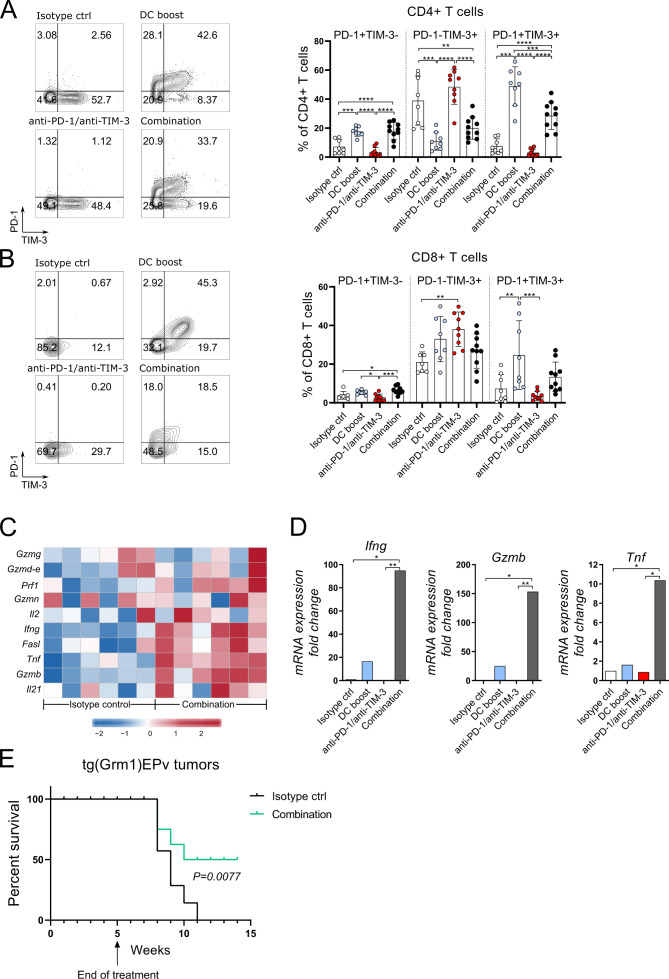
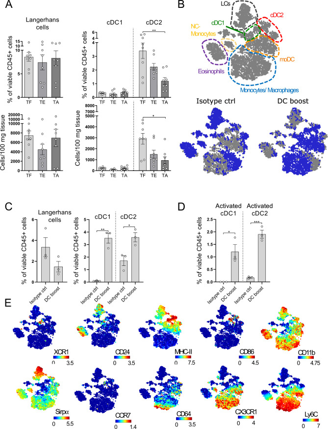
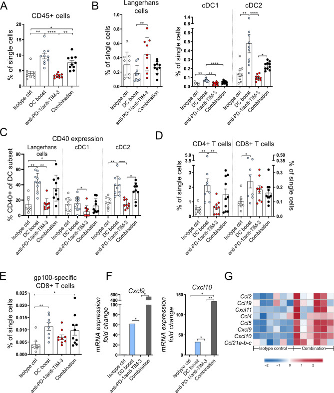
Our study revealed that tumor progression is characterized by upregulation of checkpoint molecules and a gradual loss of the dermal conventional DC (cDC) 2 subset. Monotherapy with checkpoint blockade could not restore antitumor immunity, whereas boosting DC numbers and activation increased tumor immunogenicity. This was reflected by higher numbers of activated cDC1 and cDC2 as well as CD4+ and CD8+ T cells in treated tumors. At the same time, the DC boost approach reinforced migratory dermal DC subsets to prime gp100-specific CD8+ T cells in tumor-draining LNs that expressed PD-1/TIM-3 and produced interferon γ (IFNγ)/tumor necrosis factor α (TNFα). As a consequence, the combination of the DC boost with antibodies against PD-1 and TIM-3 released the brake from T cells, leading to improved function within the tumors and delayed tumor growth.
Our results set forth the importance of skin DC in cancer immunotherapy, and demonstrates that restoring DC function is key to enhancing tumor immunogenicity and subsequently responsiveness to checkpoint blockade therapy.
免疫检查点抑制剂在黑色素瘤患者中显示出令人印象深刻的疗效,但仍有许多患者无法从中受益。肿瘤浸润 T 细胞的缺乏是治疗失败的常见原因,但也有报道称肿瘤内树突状细胞(DC)的丢失。
我们使用自发形成、生长缓慢的转基因 tg(Grm1)EPv 黑色素瘤小鼠模型,在肿瘤进展过程中进行免疫学分析。通过流式细胞术分析不同肿瘤阶段 DC 和 T 细胞的频率,以及 T 细胞上程序性死亡蛋白-1(PD-1)和 T 细胞免疫球蛋白和粘蛋白结构域 3(TIM-3)的抑制分子的表达。此外,我们还进行了 RNA 测序(RNA-seq)和实时定量 PCR(RT-qPCR)分析,以研究肿瘤的免疫状态。为了增加 DC 数量和功能,我们给予 Fms 相关酪氨酸 3 配体(Flt3L)和聚肌苷酸:聚胞苷酸与抗 CD40 的混合物作为佐剂。为了增强 T 细胞的功能,我们测试了几种检查点阻断抗体。通过流式细胞术、CyTOF、微阵列和 RT-qPCR 分析肿瘤和肿瘤引流淋巴结(LN)中的免疫改变,以了解免疫细胞如何控制肿瘤生长。通过与黑色素瘤特异性 CD8+T 细胞共培养,研究了迁移性皮肤 DC 的特定作用。
我们的研究表明,肿瘤进展的特征是检查点分子的上调和真皮常规 DC(cDC)2 亚群的逐渐丢失。单独使用检查点阻断治疗不能恢复抗肿瘤免疫,而增加 DC 数量和激活可提高肿瘤的免疫原性。这反映在治疗肿瘤中激活的 cDC1 和 cDC2 以及 CD4+和 CD8+T 细胞数量增加。与此同时,DC 促进方法增强了迁移性皮肤 DC 亚群,在肿瘤引流 LN 中引发 gp100 特异性 CD8+T 细胞,这些细胞表达 PD-1/TIM-3 并产生干扰素 γ(IFNγ)/肿瘤坏死因子 α(TNFα)。结果,DC 促进与 PD-1 和 TIM-3 的抗体联合使用,从 T 细胞中释放了制动,导致肿瘤内功能改善,并延迟了肿瘤生长。
我们的研究结果强调了皮肤 DC 在癌症免疫治疗中的重要性,并证明了恢复 DC 功能是增强肿瘤免疫原性和随后对检查点阻断治疗反应性的关键。