• 文献检索
  • 文档翻译
  • 深度研究
  • 学术资讯
  • Suppr Zotero 插件Zotero 插件
  • 邀请有礼
  • 套餐&价格
  • 历史记录
应用&插件
Suppr Zotero 插件Zotero 插件浏览器插件Mac 客户端Windows 客户端微信小程序
定价
高级版会员购买积分包购买API积分包
服务
文献检索文档翻译深度研究API 文档MCP 服务
关于我们
关于 Suppr公司介绍联系我们用户协议隐私条款
关注我们

Suppr 超能文献

核心技术专利:CN118964589B侵权必究
粤ICP备2023148730 号-1Suppr @ 2026

文献检索

告别复杂PubMed语法,用中文像聊天一样搜索,搜遍4000万医学文献。AI智能推荐,让科研检索更轻松。

立即免费搜索

文件翻译

保留排版,准确专业,支持PDF/Word/PPT等文件格式,支持 12+语言互译。

免费翻译文档

深度研究

AI帮你快速写综述,25分钟生成高质量综述,智能提取关键信息,辅助科研写作。

立即免费体验

未破裂颅内动脉瘤瘤壁增强:一项组织病理学评估。

Aneurysm Wall Enhancement in Unruptured Intracranial Aneurysms: A Histopathological Evaluation.

机构信息

Department of Neurosurgery Qilu Hospital of Shandong University and Institute of Brain and Brain-Inspired ScienceShandong University Jinan Shandong Province China.

Shandong Key Laboratory of Brain Function Remodeling Jinan Shandong Province China.

出版信息

J Am Heart Assoc. 2021 Jan 19;10(2):e018633. doi: 10.1161/JAHA.120.018633. Epub 2021 Jan 7.

DOI:10.1161/JAHA.120.018633
PMID:33410330
原文链接:https://pmc.ncbi.nlm.nih.gov/articles/PMC7955308/
Abstract

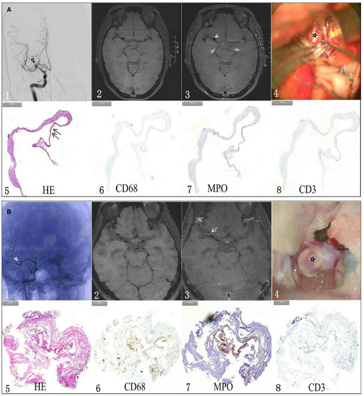
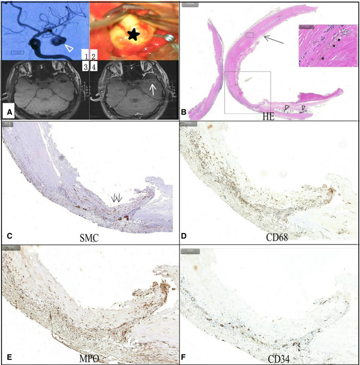
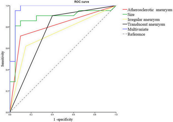
Background Unruptured intracerebral aneurysm wall enhancement (AWE) on vessel wall magnetic resonance imaging scans may be a promising predictor for rupture-prone intracerebral aneurysms. However, the pathophysiology of AWE remains unclear. To this end, the association between AWE and histopathological changes was assessed in this study. Methods and Results A total of 35 patients with 41 unruptured intracerebral aneurysms who underwent surgical clipping were prospectively enrolled. A total of 27 aneurysms were available for histological evaluation. The macroscopic and microscopic features of unruptured intracerebral aneurysms with and without enhancement were assessed. The microscopic features studied included inflammatory cell invasion and vasa vasorum, which were assessed using immunohistochemical staining with CD68, CD3, CD20, and myeloperoxidase for the former and CD34 for the latter. A total of 21 (51.2%) aneurysms showed AWE (partial AWE, n=7; circumferential AWE, n=14). Atherosclerotic and translucent aneurysms were identified in 17 and 14 aneurysms, respectively. Aneurysm size, irregularity, and atherosclerotic and translucent aneurysms were associated with AWE on univariate analysis (<0.05). Multivariate logistic regression analysis showed that atherosclerosis was the only factor significantly and independently associated with AWE (=0.027). Histological assessment revealed that inflammatory cell infiltration, intraluminal thrombus, and vasa vasorum were significantly associated with AWE (<0.05). Conclusions Though AWE on vessel wall magnetic resonance imaging scans may be associated with the presence of atherosclerotic lesions in unruptured intracerebral aneurysms, inflammatory cell infiltration within atherosclerosis, intraluminal thrombus, and vasa vasorum may be the main pathological features associated with AWE. However, the underlying pathological mechanism for AWE still needs to be further studied.

摘要

背景

血管壁磁共振成像扫描显示的未破裂颅内动脉瘤壁增强(AWE)可能是预测易破裂颅内动脉瘤的有前途的指标。然而,AWE 的病理生理学仍然不清楚。为此,本研究评估了 AWE 与组织病理学变化之间的关系。

方法和结果

本研究前瞻性纳入了 35 名接受手术夹闭的 41 个未破裂颅内动脉瘤患者。共有 27 个动脉瘤可用于组织学评估。评估了增强和未增强的未破裂颅内动脉瘤的大体和微观特征。研究的微观特征包括炎症细胞浸润和血管周腔,使用 CD68、CD3、CD20 和髓过氧化物酶对前者进行免疫组织化学染色,并用 CD34 对后者进行免疫组织化学染色。共有 21 个(51.2%)动脉瘤显示 AWE(部分 AWE,n=7;环形 AWE,n=14)。分别在 17 个和 14 个动脉瘤中发现了动脉粥样硬化和半透明动脉瘤。单因素分析显示,动脉瘤大小、不规则性以及动脉粥样硬化和半透明动脉瘤与 AWE 相关(<0.05)。多因素逻辑回归分析显示,动脉粥样硬化是唯一与 AWE 显著相关的因素(=0.027)。组织学评估显示,炎症细胞浸润、管腔内血栓和血管周腔与 AWE 显著相关(<0.05)。

结论

尽管血管壁磁共振成像扫描上的 AWE 可能与未破裂颅内动脉瘤中的动脉粥样硬化病变有关,但动脉粥样硬化内的炎症细胞浸润、管腔内血栓和血管周腔可能是与 AWE 相关的主要病理特征。然而,AWE 的潜在病理机制仍需要进一步研究。

https://cdn.ncbi.nlm.nih.gov/pmc/blobs/5fde/7955308/42224a60c6f2/JAH3-10-e018633-g004.jpg
https://cdn.ncbi.nlm.nih.gov/pmc/blobs/5fde/7955308/7f19dc939247/JAH3-10-e018633-g001.jpg
https://cdn.ncbi.nlm.nih.gov/pmc/blobs/5fde/7955308/628cce0744ee/JAH3-10-e018633-g002.jpg
https://cdn.ncbi.nlm.nih.gov/pmc/blobs/5fde/7955308/be363f3f39b1/JAH3-10-e018633-g003.jpg
https://cdn.ncbi.nlm.nih.gov/pmc/blobs/5fde/7955308/42224a60c6f2/JAH3-10-e018633-g004.jpg

相似文献

1
Aneurysm Wall Enhancement in Unruptured Intracranial Aneurysms: A Histopathological Evaluation.未破裂颅内动脉瘤瘤壁增强:一项组织病理学评估。
J Am Heart Assoc. 2021 Jan 19;10(2):e018633. doi: 10.1161/JAHA.120.018633. Epub 2021 Jan 7.
2
Vessel Wall Enhancement in Unruptured Intracranial Aneurysms: An Indicator for Higher Risk of Rupture? High-Resolution MR Imaging and Correlated Histologic Findings.颅内未破裂动脉瘤的血管壁增强:破裂风险更高的指标?高分辨率磁共振成像及相关组织学发现。
AJNR Am J Neuroradiol. 2018 Sep;39(9):1617-1621. doi: 10.3174/ajnr.A5731. Epub 2018 Jul 19.
3
Relationship between aneurysm wall enhancement and conventional risk factors in patients with unruptured intracranial aneurysms: A black-blood MRI study.未破裂颅内动脉瘤患者动脉瘤壁强化与传统危险因素的关系:一项黑血MRI研究
Interv Neuroradiol. 2016 Oct;22(5):501-5. doi: 10.1177/1591019916653252. Epub 2016 Jun 24.
4
Relationship Between Aneurysm Wall Enhancement in Vessel Wall Magnetic Resonance Imaging and Rupture Risk of Unruptured Intracranial Aneurysms.颅内未破裂动脉瘤瘤壁增强与破裂风险的关系:血管壁磁共振成像研究
Neurosurgery. 2019 Jun 1;84(6):E385-E391. doi: 10.1093/neuros/nyy310.
5
Focal Aneurysm Wall Enhancement on Magnetic Resonance Imaging Indicates Intraluminal Thrombus and the Rupture Point.磁共振成像上的局灶性动脉瘤壁增强提示管腔内血栓和破裂点。
World Neurosurg. 2019 Jul;127:e578-e584. doi: 10.1016/j.wneu.2019.03.209. Epub 2019 Mar 28.
6
The Clinical and Morphologic Features Related to Aneurysm Wall Enhancement and Enhancement Pattern in Patients with Anterior Circulation Aneurysms.前循环动脉瘤患者的瘤壁强化相关临床及形态学特征和强化模式。
World Neurosurg. 2020 Feb;134:e649-e656. doi: 10.1016/j.wneu.2019.10.156. Epub 2019 Nov 2.
7
Vessel Wall Imaging Predicts the Presence of Atherosclerotic Lesions in Unruptured Intracranial Aneurysms.血管壁成像可预测未破裂颅内动脉瘤中的动脉粥样硬化病变。
World Neurosurg. 2019 Dec;132:e775-e782. doi: 10.1016/j.wneu.2019.08.019. Epub 2019 Aug 12.
8
Qualitative and Quantitative Wall Enhancement on Magnetic Resonance Imaging Is Associated With Symptoms of Unruptured Intracranial Aneurysms.磁共振成像上的定性和定量壁增强与未破裂颅内动脉瘤的症状有关。
Stroke. 2021 Jan;52(1):213-222. doi: 10.1161/STROKEAHA.120.029685. Epub 2020 Dec 22.
9
Decreased Antiatherogenic Protein Levels are Associated with Aneurysm Structure Alterations in MR Vessel Wall Imaging.抗动脉粥样硬化蛋白水平降低与磁共振血管壁成像中动脉瘤结构改变相关。
J Stroke Cerebrovasc Dis. 2019 Aug;28(8):2221-2227. doi: 10.1016/j.jstrokecerebrovasdis.2019.05.002. Epub 2019 May 24.
10
Association of preoperative aneurysmal wall enhancement with relief of chronic headache after surgical clipping of unruptured intracranial aneurysms.术前动脉瘤壁强化与未破裂颅内动脉瘤夹闭术后慢性头痛缓解的相关性研究。
Eur Radiol. 2024 May;34(5):3009-3018. doi: 10.1007/s00330-023-10303-0. Epub 2023 Oct 20.

引用本文的文献

1
Aneurysm Wall Enhancement and Systemic Inflammation Jointly Contribute to Cognitive Dysfunction in Untreated Unruptured Intracranial Aneurysm Patients.动脉瘤壁强化与全身炎症共同导致未治疗的未破裂颅内动脉瘤患者出现认知功能障碍。
J Inflamm Res. 2025 Jul 10;18:9045-9053. doi: 10.2147/JIR.S515856. eCollection 2025.
2
Comprehensive morphomechanical and wall enhancement analysis of intracranial aneurysms.颅内动脉瘤的综合形态力学及壁强化分析
Eur Radiol. 2025 Feb 13. doi: 10.1007/s00330-025-11417-3.
3
Enhancing intracranial aneurysm rupture risk prediction with a novel multivariable logistic regression model incorporating high-resolution vessel wall imaging.

本文引用的文献

1
Treatment and diagnosis of cerebral aneurysms in the post-International Subarachnoid Aneurysm Trial (ISAT) era: trends and outcomes.国际蛛网膜下腔动脉瘤试验(ISAT)后时代脑动脉瘤的治疗和诊断:趋势和结果。
J Neurointerv Surg. 2020 Jul;12(7):682-687. doi: 10.1136/neurintsurg-2019-015418. Epub 2020 Jan 20.
2
Vessel wall magnetic resonance imaging in intracranial aneurysms: Principles and emerging clinical applications.颅内动脉瘤的管壁磁共振成像:原理及新兴临床应用。
Interv Neuroradiol. 2020 Apr;26(2):135-146. doi: 10.1177/1591019919891297. Epub 2019 Dec 9.
3
The Clinical and Morphologic Features Related to Aneurysm Wall Enhancement and Enhancement Pattern in Patients with Anterior Circulation Aneurysms.
利用纳入高分辨率血管壁成像的新型多变量逻辑回归模型增强颅内动脉瘤破裂风险预测
Front Neurol. 2025 Jan 7;15:1507082. doi: 10.3389/fneur.2024.1507082. eCollection 2024.
4
Irregular pulsation on 4D-CTA and quantitative wall enhancement on VW-MRI are associated with symptoms of unruptured intracranial aneurysms.4D-CTA上的不规则搏动和VW-MRI上的定量壁强化与未破裂颅内动脉瘤的症状相关。
J Neurointerv Surg. 2025 Jan 27. doi: 10.1136/jnis-2024-022483.
5
A multiscale discrete fiber model of failure in heterogeneous tissues: Applications to remodeled cerebral aneurysms.异质组织失效的多尺度离散纤维模型:在重塑脑动脉瘤中的应用。
J Biomech. 2025 Jan;178:112343. doi: 10.1016/j.jbiomech.2024.112343. Epub 2024 Sep 24.
6
There Is Poor Agreement between the Subjective and Quantitative Adjudication of Aneurysm Wall Enhancement.动脉瘤壁强化的主观判定与定量判定之间的一致性较差。
AJNR Am J Neuroradiol. 2025 Apr 2;46(4):689-697. doi: 10.3174/ajnr.A8508.
7
Vasa vasorum: The role in intracranial physiology and pathophysiology.血管滋养血管:在颅内生理学和病理生理学中的作用
Surg Neurol Int. 2024 Jun 7;15:188. doi: 10.25259/SNI_214_2024. eCollection 2024.
8
Imaging of Intracranial Aneurysms: A Review of Standard and Advanced Imaging Techniques.颅内动脉瘤的影像学检查:标准及先进成像技术综述
Transl Stroke Res. 2025 Jun;16(3):1016-1027. doi: 10.1007/s12975-024-01261-w. Epub 2024 Jun 10.
9
Wall Enhancement of Coiled Intracranial Aneurysms Is Associated with Aneurysm Recanalization: A Cross-Sectional Study.颅内动脉瘤线圈增强与动脉瘤再通相关:一项横断面研究。
AJNR Am J Neuroradiol. 2024 May 9;45(5):599-604. doi: 10.3174/ajnr.A8174.
10
A Systematic Review and Meta-Analysis of the Pathology Underlying Aneurysm Enhancement on Vessel Wall Imaging.血管壁成像中动脉瘤强化的病理基础的系统评价和荟萃分析。
Int J Mol Sci. 2024 Feb 26;25(5):2700. doi: 10.3390/ijms25052700.
前循环动脉瘤患者的瘤壁强化相关临床及形态学特征和强化模式。
World Neurosurg. 2020 Feb;134:e649-e656. doi: 10.1016/j.wneu.2019.10.156. Epub 2019 Nov 2.
4
Vessel Wall Imaging Predicts the Presence of Atherosclerotic Lesions in Unruptured Intracranial Aneurysms.血管壁成像可预测未破裂颅内动脉瘤中的动脉粥样硬化病变。
World Neurosurg. 2019 Dec;132:e775-e782. doi: 10.1016/j.wneu.2019.08.019. Epub 2019 Aug 12.
5
Gadolinium Enhancement of the Aneurysm Wall in Unruptured Intracranial Aneurysms Is Associated with an Increased Risk of Aneurysm Instability: A Follow-Up Study.未破裂颅内动脉瘤瘤壁钆增强与动脉瘤不稳定风险增加相关:一项随访研究。
AJNR Am J Neuroradiol. 2019 Jul;40(7):1112-1116. doi: 10.3174/ajnr.A6105. Epub 2019 Jun 20.
6
Vessel Wall Imaging of Evolving Unruptured Intracranial Aneurysms.颅内未破裂动脉瘤演变的血管壁成像。
Stroke. 2019 Jul;50(7):1891-1894. doi: 10.1161/STROKEAHA.119.025245. Epub 2019 Jun 6.
7
Wall Contrast Enhancement of Thrombosed Intracranial Aneurysms at 7T MRI.7T MRI 下血栓形成颅内动脉瘤的壁对比度增强。
AJNR Am J Neuroradiol. 2019 Jul;40(7):1106-1111. doi: 10.3174/ajnr.A6084. Epub 2019 May 30.
8
Validation of Wall Enhancement as a New Imaging Biomarker of Unruptured Cerebral Aneurysm.颅内未破裂动脉瘤的壁强化作为一种新的成像生物标志物的验证。
Stroke. 2019 Jun;50(6):1570-1573. doi: 10.1161/STROKEAHA.118.024195. Epub 2019 Apr 30.
9
Magnetic Resonance Vessel Wall Imaging in Human Intracranial Aneurysms: Histological Analysis.人体颅内动脉瘤的磁共振血管壁成像:组织学分析
Stroke. 2019 Jan;50(1):e1. doi: 10.1161/STROKEAHA.118.023701. Epub 2018 Dec 7.
10
Clinicopathological Insights From Vessel Wall Imaging of Unruptured Intracranial Aneurysms.未破裂颅内动脉瘤血管壁影像学的临床病理见解。
Stroke. 2018 Oct;49(10):2516-2519. doi: 10.1161/STROKEAHA.118.021819.