• 文献检索
  • 文档翻译
  • 深度研究
  • 学术资讯
  • Suppr Zotero 插件Zotero 插件
  • 邀请有礼
  • 套餐&价格
  • 历史记录
应用&插件
Suppr Zotero 插件Zotero 插件浏览器插件Mac 客户端Windows 客户端微信小程序
定价
高级版会员购买积分包购买API积分包
服务
文献检索文档翻译深度研究API 文档MCP 服务
关于我们
关于 Suppr公司介绍联系我们用户协议隐私条款
关注我们

Suppr 超能文献

核心技术专利:CN118964589B侵权必究
粤ICP备2023148730 号-1Suppr @ 2026

文献检索

告别复杂PubMed语法,用中文像聊天一样搜索,搜遍4000万医学文献。AI智能推荐,让科研检索更轻松。

立即免费搜索

文件翻译

保留排版,准确专业,支持PDF/Word/PPT等文件格式,支持 12+语言互译。

免费翻译文档

深度研究

AI帮你快速写综述,25分钟生成高质量综述,智能提取关键信息,辅助科研写作。

立即免费体验

全球光学相干断层扫描测量在检测青光眼进展方面存在根本缺陷。

Global optical coherence tomography measures for detecting the progression of glaucoma have fundamental flaws.

机构信息

Department of Psychology, Columbia University, New York, NY, USA.

Einhorn Clinical Research Center, New York Eye and Ear Infirmary of Mount Sinai, New York, NY, USA.

出版信息

Eye (Lond). 2021 Nov;35(11):2973-2982. doi: 10.1038/s41433-020-01296-x. Epub 2021 Jan 7.

DOI:10.1038/s41433-020-01296-x
PMID:33414534
原文链接:https://pmc.ncbi.nlm.nih.gov/articles/PMC8526823/
Abstract

OBJECTIVE

To understand the problems involved in using global OCT measures for detecting progression in early glaucoma.

SUBJECTS/METHODS: Eyes from 76 patients and 28 healthy controls (HC) had a least two OCT scans at least 1 year apart. To determine the 95% confidence intervals (CI), 151 eyes (49 HC and 102 patients) had at least two scans within 6 months. All eyes had 24-2 mean deviation ≥-6dB. The average (global) thicknesses of the circumpapillary retinal nerve fibre layer (cRNFL), G, and of the retinal ganglion cell layer plus inner plexiform layer (RGCLP), G, were calculated. Using quantile regression, the 95% CI intervals were determined. Eyes outside the CIs were classified as "progressors." For a reference standard (RS), four experts evaluated OCT and VF information.

RESULTS

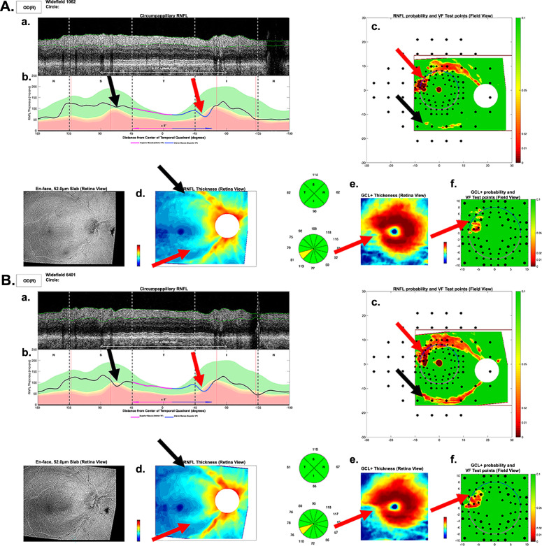
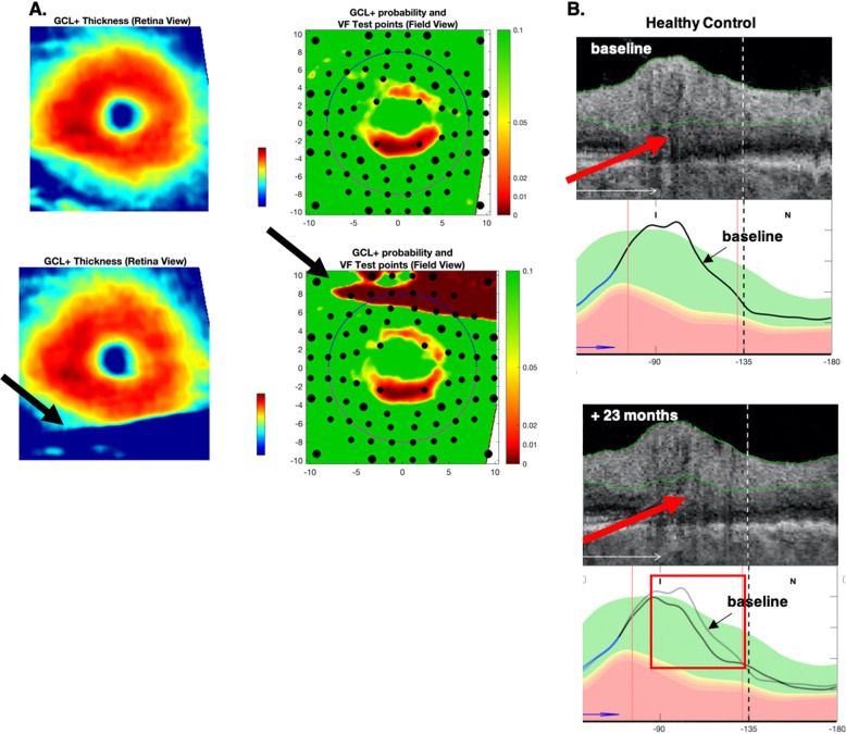
Compared to the RS, 31 of the 76 (40.8%) patient eyes were identified as progressors (RS-P), and 45 patient, and all 28 HC, eyes as nonprogressors (RS-NP). The metrics missed (false negative, FN) 15 (48%) (G) and 9 (29%) (G) of the 31 RS-P. Further, G and/or G falsely identified (false positive, FP) 10 (22.2%) of 45 patient RS-NP eyes and 7 (25%) of the 28 HC eyes as progressing. Post-hoc analysis identified three reasons (segmentation, centring, and local damage) for these errors.

CONCLUSIONS

Global metrics lead to FPs and FNs because of problems inherent in OCT scanning (segmentation and centring), and to FNs because they can miss local damage. These problems are difficult, if not impossible, to correct, and raise concerns about the advisability of using G and G for detecting progression.

摘要

目的

了解使用全球 OCT 测量值检测早期青光眼进展所涉及的问题。

对象/方法:76 名患者和 28 名健康对照者(HC)的眼睛至少相隔 1 年进行了两次 OCT 扫描。为了确定 95%置信区间(CI),151 只眼睛(49 只 HC 和 102 只患者)在 6 个月内至少进行了两次扫描。所有眼睛的 24-2 平均偏差(MD)≥-6dB。计算了环周视网膜神经纤维层(cRNFL)、G 和视网膜神经节细胞层加内丛状层(RGCLP)、G 的平均(整体)厚度。使用分位数回归确定 95%CI 间隔。超出 CI 的眼睛被分类为“进展者”。对于参考标准(RS),四位专家评估了 OCT 和 VF 信息。

结果

与 RS 相比,76 名患者中有 31 只(40.8%)眼睛被确定为进展者(RS-P),45 名患者和所有 28 名 HC 眼睛被确定为非进展者(RS-NP)。指标遗漏(假阴性,FN)了 31 个 RS-P 中的 15 个(G)和 9 个(G)。此外,G 和/或 G 错误地将 45 名患者 RS-NP 中的 10 只(22.2%)和 28 名 HC 中的 7 只(25%)眼睛识别为进展。事后分析确定了这些错误的三个原因(分割、居中、和局部损伤)。

结论

由于 OCT 扫描(分割和居中)固有的问题,全局指标会导致 FP 和 FN,并因遗漏局部损伤而导致 FN。这些问题很难(如果不是不可能)纠正,这引发了对使用 G 和 G 检测进展的合理性的担忧。

https://cdn.ncbi.nlm.nih.gov/pmc/blobs/97fc/8526823/ed9fd261f70c/41433_2020_1296_Fig4_HTML.jpg
https://cdn.ncbi.nlm.nih.gov/pmc/blobs/97fc/8526823/019c7fb9d84c/41433_2020_1296_Fig1_HTML.jpg
https://cdn.ncbi.nlm.nih.gov/pmc/blobs/97fc/8526823/18ad2311c75a/41433_2020_1296_Fig2_HTML.jpg
https://cdn.ncbi.nlm.nih.gov/pmc/blobs/97fc/8526823/e94f5d102d55/41433_2020_1296_Fig3_HTML.jpg
https://cdn.ncbi.nlm.nih.gov/pmc/blobs/97fc/8526823/ed9fd261f70c/41433_2020_1296_Fig4_HTML.jpg

相似文献

1
Global optical coherence tomography measures for detecting the progression of glaucoma have fundamental flaws.全球光学相干断层扫描测量在检测青光眼进展方面存在根本缺陷。
Eye (Lond). 2021 Nov;35(11):2973-2982. doi: 10.1038/s41433-020-01296-x. Epub 2021 Jan 7.
2
Ganglion Cell-Inner Plexiform Layer Change Detected by Optical Coherence Tomography Indicates Progression in Advanced Glaucoma.光学相干断层扫描检测到的神经节细胞-内丛状层变化表明晚期青光眼的进展。
Ophthalmology. 2017 Oct;124(10):1466-1474. doi: 10.1016/j.ophtha.2017.04.023. Epub 2017 May 23.
3
The 24-2 Visual Field Guided Progression Analysis Can Miss the Progression of Glaucomatous Damage of the Macula Seen Using OCT.24-2 视野引导进展分析可能会错过 OCT 观察到的黄斑青光眼损伤的进展。
Ophthalmol Glaucoma. 2022 Nov-Dec;5(6):614-627. doi: 10.1016/j.ogla.2022.03.007. Epub 2022 Mar 28.
4
Detection of glaucomatous progression by spectral-domain optical coherence tomography.利用频域光学相干断层扫描检测青光眼进展。
Ophthalmology. 2013 Jul;120(7):1388-95. doi: 10.1016/j.ophtha.2012.12.014. Epub 2013 Mar 6.
5
Detecting Progression in Advanced Glaucoma: Are Optical Coherence Tomography Global Metrics Viable Measures?检测晚期青光眼的进展:光学相干断层扫描全局指标是否可行的测量方法?
Optom Vis Sci. 2021 May 1;98(5):518-530. doi: 10.1097/OPX.0000000000001697.
6
Detecting Glaucoma Progression Using Guided Progression Analysis with OCT and Visual Field Assessment in Eyes Classified by International Classification of Disease Severity Codes.基于国际疾病严重程度分类代码对眼部进行分类,利用 OCT 和视野评估的引导进展分析来检测青光眼进展。
Ophthalmol Glaucoma. 2019 Jan-Feb;2(1):36-46. doi: 10.1016/j.ogla.2018.11.004. Epub 2018 Nov 15.
7
Association Between Progressive Retinal Capillary Density Loss and Visual Field Progression in Open-Angle Glaucoma Patients According to Disease Stage.根据疾病阶段,广角型青光眼患者视网膜毛细血管密度进行性损失与视野进展之间的关系。
Am J Ophthalmol. 2021 Jun;226:137-147. doi: 10.1016/j.ajo.2021.01.015. Epub 2021 Jan 30.
8
Progression of Early Glaucomatous Damage: Performance of Summary Statistics From Optical Coherence Tomography and Perimetry.早期青光眼损害的进展:光学相干断层扫描和视野计汇总统计数据的表现。
Transl Vis Sci Technol. 2023 Mar 1;12(3):19. doi: 10.1167/tvst.12.3.19.
9
Trend-based Analysis of Ganglion Cell-Inner Plexiform Layer Thickness Changes on Optical Coherence Tomography in Glaucoma Progression.基于趋势的青光眼进展中光学相干断层扫描的神经节细胞-内丛状层厚度变化分析。
Ophthalmology. 2017 Sep;124(9):1383-1391. doi: 10.1016/j.ophtha.2017.03.013. Epub 2017 Apr 12.
10
Wide-field Trend-based Progression Analysis of Combined Retinal Nerve Fiber Layer and Ganglion Cell Inner Plexiform Layer Thickness: A New Paradigm to Improve Glaucoma Progression Detection.基于广角趋势的视网膜神经纤维层和节细胞内丛状层联合厚度进展分析:一种提高青光眼进展检测的新方法。
Ophthalmology. 2020 Oct;127(10):1322-1330. doi: 10.1016/j.ophtha.2020.03.019. Epub 2020 Mar 29.

引用本文的文献

1
A Pattern-Based OCT Metric for Glaucoma Detection.一种基于模式的用于青光眼检测的光学相干断层扫描指标。
Transl Vis Sci Technol. 2024 Dec 2;13(12):21. doi: 10.1167/tvst.13.12.21.
2
A Model of Progression to Help Identify Macular Damage Due to Glaucoma.有助于识别青光眼导致的黄斑损伤的进展模型。
Invest Ophthalmol Vis Sci. 2023 Dec 1;64(15):8. doi: 10.1167/iovs.64.15.8.
3
Progression of Early Glaucomatous Damage: Performance of Summary Statistics From Optical Coherence Tomography and Perimetry.早期青光眼损害的进展:光学相干断层扫描和视野计汇总统计数据的表现。
Transl Vis Sci Technol. 2023 Mar 1;12(3):19. doi: 10.1167/tvst.12.3.19.
4
Prediction of visual field defects from macular optical coherence tomography in glaucoma using cluster analysis.利用聚类分析从青光眼的黄斑光学相干断层扫描中预测视野缺损。
Ophthalmic Physiol Opt. 2022 Sep;42(5):948-964. doi: 10.1111/opo.12997. Epub 2022 May 22.
5
Detection of Early Glaucomatous Damage: Performance of Summary Statistics From Optical Coherence Tomography and Perimetry.早期青光眼损害的检测:光学相干断层扫描和视野计汇总统计数据的性能。
Transl Vis Sci Technol. 2022 Mar 2;11(3):36. doi: 10.1167/tvst.11.3.36.
6
The OCT RNFL Probability Map and Artifacts Resembling Glaucomatous Damage.OCT 神经纤维层概率图与青光眼损害类似的伪影。
Transl Vis Sci Technol. 2022 Mar 2;11(3):18. doi: 10.1167/tvst.11.3.18.
7
Rationale and Development of an OCT-Based Method for Detection of Glaucomatous Optic Neuropathy.基于 OCT 的青光眼视神经病变检测方法的原理与发展。
J Glaucoma. 2022 Jun 1;31(6):375-381. doi: 10.1097/IJG.0000000000002005. Epub 2022 Feb 28.
8
Did the OCT Show Progression Since the Last Visit?OCT 显示自上次就诊以来是否有进展?
J Glaucoma. 2021 Apr 1;30(4):e134-e145. doi: 10.1097/IJG.0000000000001766.
9
Reasons why OCT Global Circumpapillary Retinal Nerve Fiber Layer Thickness is a Poor Measure of Glaucomatous Progression.光学相干断层扫描(OCT)测量的全球视盘周围视网膜神经纤维层厚度为何不是评估青光眼病情进展的理想指标。
Transl Vis Sci Technol. 2020 Oct 19;9(11):22. doi: 10.1167/tvst.9.11.22. eCollection 2020 Oct.