• 文献检索
  • 文档翻译
  • 深度研究
  • 学术资讯
  • Suppr Zotero 插件Zotero 插件
  • 邀请有礼
  • 套餐&价格
  • 历史记录
应用&插件
Suppr Zotero 插件Zotero 插件浏览器插件Mac 客户端Windows 客户端微信小程序
定价
高级版会员购买积分包购买API积分包
服务
文献检索文档翻译深度研究API 文档MCP 服务
关于我们
关于 Suppr公司介绍联系我们用户协议隐私条款
关注我们

Suppr 超能文献

核心技术专利:CN118964589B侵权必究
粤ICP备2023148730 号-1Suppr @ 2026

文献检索

告别复杂PubMed语法,用中文像聊天一样搜索,搜遍4000万医学文献。AI智能推荐,让科研检索更轻松。

立即免费搜索

文件翻译

保留排版,准确专业,支持PDF/Word/PPT等文件格式,支持 12+语言互译。

免费翻译文档

深度研究

AI帮你快速写综述,25分钟生成高质量综述,智能提取关键信息,辅助科研写作。

立即免费体验

OCT 神经纤维层概率图与青光眼损害类似的伪影。

The OCT RNFL Probability Map and Artifacts Resembling Glaucomatous Damage.

机构信息

Department of Psychology, Columbia University, New York, NY, USA.

Albert Einstein College of Medicine, New York, NY, USA.

出版信息

Transl Vis Sci Technol. 2022 Mar 2;11(3):18. doi: 10.1167/tvst.11.3.18.

DOI:10.1167/tvst.11.3.18
PMID:35289836
原文链接:https://pmc.ncbi.nlm.nih.gov/articles/PMC8934545/
Abstract

PURPOSE

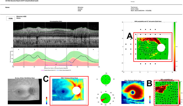
The purpose of this study was to improve the diagnostic ability of the optical coherence tomography (OCT) retinal nerve fiber layer (RNFL) probability (p-) map by understanding the frequency and pattern of artifacts seen on the p-maps of healthy control (HC) eyes resembling glaucomatous damage.

METHODS

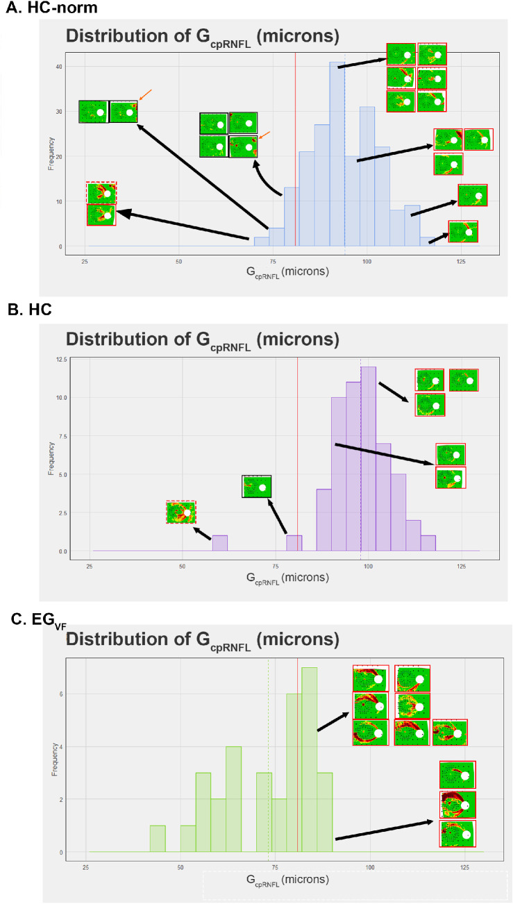
RNFL p-maps were generated from wide-field OCT cube scans of 2 groups of HC eyes, 200 from a commercial normative group (HC-norm) and 54 from a prospective study group, as well as from 62 patient eyes, which included 32 with early glaucoma (EG). These 32 EG eyes had 24-2 mean deviation (MD) better than -6 dB and perimetric glaucoma as defined by 24-2 and 10-2 criteria. For the HC groups, "glaucoma-like" arcuates were defined as any red region near the temporal half of the disc.

RESULTS

Seven percent of the 200 HC-norm and 11% of the 54 HC RNFL p-maps satisfied the definition of "glaucoma-like," as did all the patients' p-maps. The HC p-maps showed two general patterns of abnormal regions, "arcuate" and "temporal quadrant," and these patterns resembled those seen on some of the RNFL p-maps of the EG eyes. A "vertical midline" rule, which required the abnormal region to cross the vertical midline through the fovea, had a specificity of >99%, and a sensitivity of 75% for EG and 93% for moderate to advanced eyes.

CONCLUSIONS

Glaucoma-like artifacts on RNFL p-maps are relatively common and can masquerade as arcuate and/or widespread/temporal damage.

TRANSLATIONAL RELEVANCE

A vertical midline rule had excellent specificity. However, other OCT information is necessary to obtain high sensitivity, especially in eyes with early glaucoma.

摘要

目的

本研究旨在通过了解类似于青光眼损伤的健康对照(HC)眼 OCT 视网膜神经纤维层(RNFL)概率(p-)图中出现的伪影的频率和模式,提高 OCT 视网膜神经纤维层(RNFL)概率(p-)图的诊断能力。

方法

从两组 HC 眼的广角 OCT 立方体扫描中生成 RNFL p-图,一组来自商业正常组(HC-norm)的 200 只眼,另一组来自前瞻性研究组的 54 只眼,以及 62 只患者眼,其中包括 32 只早期青光眼(EG)眼。这 32 只 EG 眼的 24-2 平均偏差(MD)比-6 dB 好,并且根据 24-2 和 10-2 标准被认为是周边青光眼。对于 HC 组,“青光眼样”弓形定义为视盘颞半部分附近的任何红色区域。

结果

200 只 HC-norm 中有 7%和 54 只 HC RNFL p-图符合“青光眼样”的定义,所有患者的 p-图也符合。HC p-图显示了两种异常区域的一般模式,“弓形”和“颞象限”,这些模式类似于一些 EG 眼的 RNFL p-图上看到的模式。“垂直中线”规则要求异常区域通过黄斑穿过垂直中线,其特异性>99%,对 EG 眼的敏感性为 75%,对中度至晚期眼的敏感性为 93%。

结论

RNFL p-图上的青光眼样伪影相对常见,可能伪装为弓形和/或广泛/颞部损伤。

翻译

医学

https://cdn.ncbi.nlm.nih.gov/pmc/blobs/eee1/8934545/32fd2d64ae25/tvst-11-3-18-f005.jpg
https://cdn.ncbi.nlm.nih.gov/pmc/blobs/eee1/8934545/763a90f240da/tvst-11-3-18-f001.jpg
https://cdn.ncbi.nlm.nih.gov/pmc/blobs/eee1/8934545/daf73622cb54/tvst-11-3-18-f002.jpg
https://cdn.ncbi.nlm.nih.gov/pmc/blobs/eee1/8934545/737c90dfefbb/tvst-11-3-18-f003.jpg
https://cdn.ncbi.nlm.nih.gov/pmc/blobs/eee1/8934545/87526e88ad79/tvst-11-3-18-f004.jpg
https://cdn.ncbi.nlm.nih.gov/pmc/blobs/eee1/8934545/32fd2d64ae25/tvst-11-3-18-f005.jpg

相似文献

1
The OCT RNFL Probability Map and Artifacts Resembling Glaucomatous Damage.OCT 神经纤维层概率图与青光眼损害类似的伪影。
Transl Vis Sci Technol. 2022 Mar 2;11(3):18. doi: 10.1167/tvst.11.3.18.
2
Diagnostic Ability of Wide-field Retinal Nerve Fiber Layer Maps Using Swept-Source Optical Coherence Tomography for Detection of Preperimetric and Early Perimetric Glaucoma.使用扫频光学相干断层扫描技术的广角视网膜神经纤维层图谱对视野缺损前和早期视野缺损性青光眼的诊断能力
J Glaucoma. 2017 Jun;26(6):577-585. doi: 10.1097/IJG.0000000000000662.
3
Ability of cirrus high-definition spectral-domain optical coherence tomography clock-hour, deviation, and thickness maps in detecting photographic retinal nerve fiber layer abnormalities.高频谱域光相干断层扫描钟点数、偏转角和厚度图在检测照相性视网膜神经纤维层异常中的作用。
Ophthalmology. 2013 Jul;120(7):1380-7. doi: 10.1016/j.ophtha.2012.12.048. Epub 2013 Mar 28.
4
Comparison of glaucoma-diagnostic ability between wide-field swept-source OCT retinal nerve fiber layer maps and spectral-domain OCT.宽视野扫频源光学相干断层扫描视网膜神经纤维层图与谱域光学相干断层扫描诊断青光眼能力的比较。
Eye (Lond). 2018 Sep;32(9):1483-1492. doi: 10.1038/s41433-018-0104-5. Epub 2018 May 23.
5
Serial Combined Wide-Field Optical Coherence Tomography Maps for Detection of Early Glaucomatous Structural Progression.用于检测早期青光眼结构进展的串行组合广角光学相干断层扫描图。
JAMA Ophthalmol. 2018 Oct 1;136(10):1121-1127. doi: 10.1001/jamaophthalmol.2018.3160.
6
Macular ganglion cell analysis for early detection of glaucoma.黄斑神经节细胞分析用于青光眼的早期检测。
Ophthalmology. 2014 Aug;121(8):1508-15. doi: 10.1016/j.ophtha.2014.02.019. Epub 2014 Apr 3.
7
Diagnostic classification of macular ganglion cell and retinal nerve fiber layer analysis: differentiation of false-positives from glaucoma.黄斑神经节细胞和视网膜神经纤维层分析的诊断分类:从青光眼区分假阳性。
Ophthalmology. 2015 Mar;122(3):502-10. doi: 10.1016/j.ophtha.2014.09.031. Epub 2014 Nov 14.
8
Separation and thickness measurements of superficial and deep slabs of the retinal nerve fiber layer in healthy and glaucomatous eyes.健康眼和青光眼眼中视网膜神经纤维层浅层和深层板的分离和厚度测量。
Ophthalmol Glaucoma. 2020 Jan-Feb;3(1):66-75. doi: 10.1016/j.ogla.2019.11.004. Epub 2019 Nov 20.
9
Retinal nerve fiber layer imaging with spectral-domain optical coherence tomography: analysis of the retinal nerve fiber layer map for glaucoma detection.频域光学相干断层扫描的视网膜神经纤维层成像:用于青光眼检测的视网膜神经纤维层图分析。
Ophthalmology. 2010 Sep;117(9):1684-91. doi: 10.1016/j.ophtha.2010.01.026. Epub 2010 Jul 21.
10
Assessment of a Segmentation-Free Deep Learning Algorithm for Diagnosing Glaucoma From Optical Coherence Tomography Scans.基于无分割深度学习算法的光学相干断层扫描图像青光眼诊断评估。
JAMA Ophthalmol. 2020 Apr 1;138(4):333-339. doi: 10.1001/jamaophthalmol.2019.5983.

引用本文的文献

1
Anatomical Features can Affect OCT Measures Used for Clinical Decisions and Clinical Trial Endpoints.解剖学特征可能会影响用于临床决策和临床试验终点的 OCT 测量。
Transl Vis Sci Technol. 2024 Apr 2;13(4):27. doi: 10.1167/tvst.13.4.27.
2
Predicting perimetric defects from en face maps of retinal nerve fibre layer reflectance.从视网膜神经纤维层反射的正面图预测周边视野缺损。
Ophthalmic Physiol Opt. 2024 May;44(3):613-625. doi: 10.1111/opo.13289. Epub 2024 Feb 25.
3
A Model of Progression to Help Identify Macular Damage Due to Glaucoma.

本文引用的文献

1
Detection of Early Glaucomatous Damage: Performance of Summary Statistics From Optical Coherence Tomography and Perimetry.早期青光眼损害的检测:光学相干断层扫描和视野计汇总统计数据的性能。
Transl Vis Sci Technol. 2022 Mar 2;11(3):36. doi: 10.1167/tvst.11.3.36.
2
Rationale and Development of an OCT-Based Method for Detection of Glaucomatous Optic Neuropathy.基于 OCT 的青光眼视神经病变检测方法的原理与发展。
J Glaucoma. 2022 Jun 1;31(6):375-381. doi: 10.1097/IJG.0000000000002005. Epub 2022 Feb 28.
3
Detecting glaucoma with only OCT: Implications for the clinic, research, screening, and AI development.
有助于识别青光眼导致的黄斑损伤的进展模型。
Invest Ophthalmol Vis Sci. 2023 Dec 1;64(15):8. doi: 10.1167/iovs.64.15.8.
4
Neurofilament Light Chain in Aqueous Humor as a Marker of Neurodegeneration in Glaucoma.房水中神经丝轻链作为青光眼神经退行性变的标志物
Clin Ophthalmol. 2023 Aug 2;17:2209-2217. doi: 10.2147/OPTH.S417664. eCollection 2023.
5
Toward a Real-world Optical Coherence Tomography Reference Database: Optometric Practices as a Source of Healthy Eyes.迈向真实世界的光学相干断层扫描参考数据库:视光实践作为健康眼睛的来源。
Optom Vis Sci. 2023 Aug 1;100(8):499-506. doi: 10.1097/OPX.0000000000002049. Epub 2023 Jul 27.
6
Structure-function assessment in glaucoma based on perimetric sensitivity and en face optical coherence tomography images of retinal nerve fiber bundles.基于视野敏感性和视网膜神经纤维束的共焦光学相干断层扫描图像的青光眼结构-功能评估。
Sci Rep. 2023 Feb 13;13(1):2497. doi: 10.1038/s41598-023-28917-1.
7
Within-eye and between-subject variability for reflectance of the retinal nerve fibre layer.眼内和个体间视网膜神经纤维层反射率的可变性。
Ophthalmic Physiol Opt. 2022 Nov;42(6):1316-1325. doi: 10.1111/opo.13027. Epub 2022 Aug 1.
仅用 OCT 检测青光眼:对临床、研究、筛查和 AI 发展的影响。
Prog Retin Eye Res. 2022 Sep;90:101052. doi: 10.1016/j.preteyeres.2022.101052. Epub 2022 Feb 23.
4
Wide-field optical coherence tomography deviation map for early glaucoma detection.宽场光学相干断层扫描离焦图在青光眼早期检测中的应用。
Br J Ophthalmol. 2023 Jan;107(1):49-55. doi: 10.1136/bjophthalmol-2021-319509. Epub 2021 Jul 22.
5
Optical Coherence Tomography and Glaucoma.光学相干断层扫描与青光眼。
Annu Rev Vis Sci. 2021 Sep 15;7:693-726. doi: 10.1146/annurev-vision-100419-111350. Epub 2021 Jul 9.
6
Strategies to Improve Convolutional Neural Network Generalizability and Reference Standards for Glaucoma Detection From OCT Scans.提高卷积神经网络泛化能力的策略以及从 OCT 扫描中检测青光眼的参考标准。
Transl Vis Sci Technol. 2021 Apr 1;10(4):16. doi: 10.1167/tvst.10.4.16.
7
Deep Learning-based Diagnosis of Glaucoma Using Wide-field Optical Coherence Tomography Images.基于深度学习的广角光学相干断层扫描图像青光眼诊断。
J Glaucoma. 2021 Sep 1;30(9):803-812. doi: 10.1097/IJG.0000000000001885.
8
Global optical coherence tomography measures for detecting the progression of glaucoma have fundamental flaws.全球光学相干断层扫描测量在检测青光眼进展方面存在根本缺陷。
Eye (Lond). 2021 Nov;35(11):2973-2982. doi: 10.1038/s41433-020-01296-x. Epub 2021 Jan 7.
9
Robust and Interpretable Convolutional Neural Networks to Detect Glaucoma in Optical Coherence Tomography Images.稳健且可解释的卷积神经网络,用于检测光学相干断层扫描图像中的青光眼。
IEEE Trans Biomed Eng. 2021 Aug;68(8):2456-2466. doi: 10.1109/TBME.2020.3043215. Epub 2021 Jul 19.
10
Optical Coherence Tomography Can Be Used to Assess Glaucomatous Optic Nerve Damage in Most Eyes With High Myopia.光学相干断层扫描可用于评估大多数高度近视患者的青光眼视神经损伤。
J Glaucoma. 2020 Oct;29(10):833-845. doi: 10.1097/IJG.0000000000001631.