Department of Pathology and Laboratory Medicine, Indiana University School of Medicine, Indianapolis, IN, 46202, USA.
GE Research, Niskayuna, NY, 12309, USA.
Br J Cancer. 2021 Mar;124(6):1150-1159. doi: 10.1038/s41416-020-01216-6. Epub 2021 Jan 7.
There is limited knowledge about DCIS cellular composition and relationship with breast cancer events (BCE).
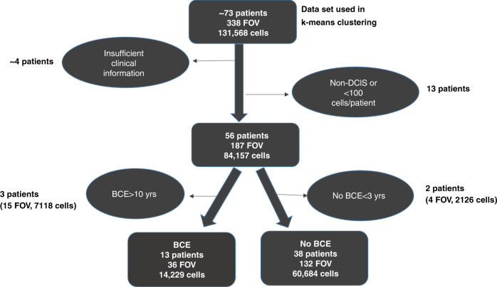
Immunofluorescence multiplexing (MxIF) was used to image and quantify 32 cellular biomarkers in FFPE DCIS tissue microarrays. Over 75,000 DCIS cells from 51 patients (median 9 years follow-up for non-BCE cases) were analysed for profiles predictive of BCE. K-means clustering was used to evaluate cellular co-expression of epithelial markers with ER and HER2.
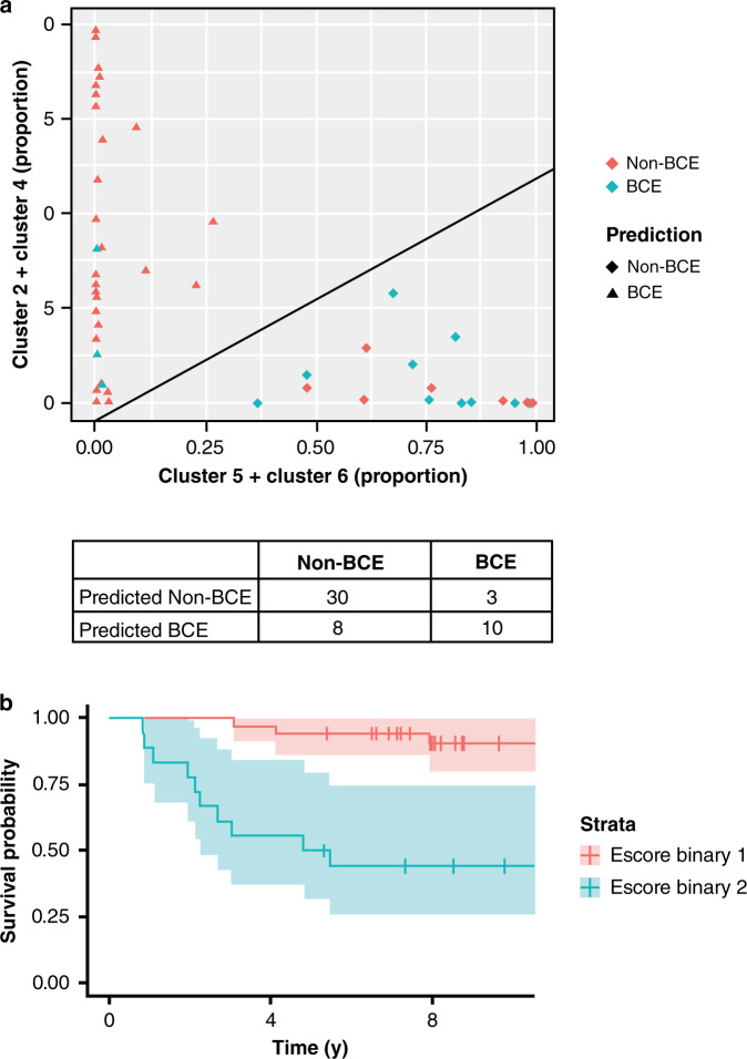
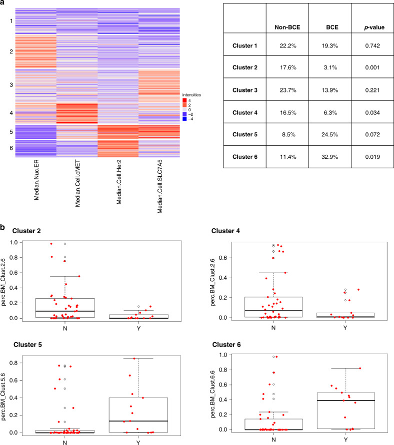
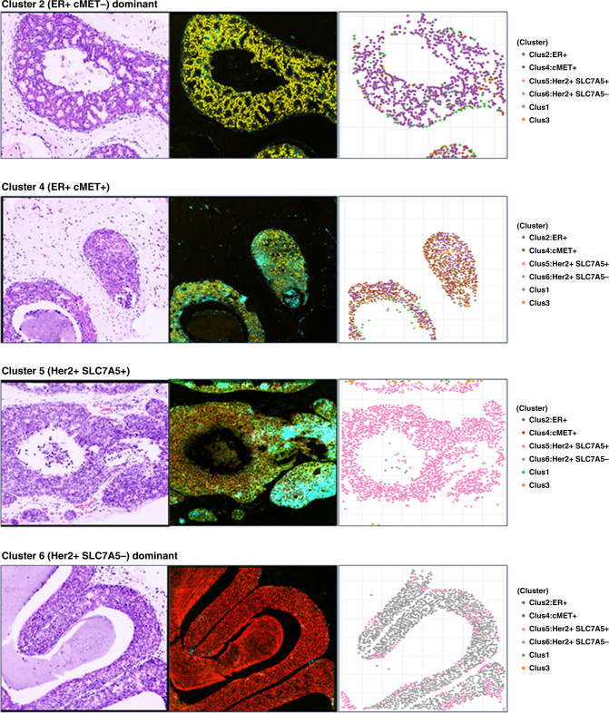
Only ER, PR and HER2 significantly correlated with BCE. Cluster analysis identified 6 distinct cell groups with different levels of ER, Her2, cMET and SLC7A5. Clusters 1 and 3 were not significant. Clusters 2 and 4 (high ER/low HER2 and SLC7A5/mixed cMET) significantly correlated with low BCE risk (P = 0.001 and P = 0.034), while cluster 6 (high HER2/low ER, cMET and SLC7A5) correlated with increased risk (P = 0.018). Cluster 5 (similar to cluster 6, except high SLC7A5) trended towards significance (P = 0.072). A continuous expression score (Escore) based on these 4 clusters predicted likelihood of BCE (AUC = 0.79, log-rank test P = 5E-05; LOOCV AUC = 0.74, log-rank test P = 0.006).
Multiplexed spatial analysis of limited tissue is a novel method for biomarker analysis and predicting BCEs. Further validation of Escore is needed in a larger cohort.
关于 DCIS 细胞组成及其与乳腺癌事件 (BCE) 的关系,目前人们的了解有限。
使用免疫荧光多重检测(MxIF)对 FFPE DCIS 组织微阵列中的 32 种细胞生物标志物进行成像和定量分析。对 51 例患者(非 BCE 病例的中位随访时间为 9 年)的超过 75000 个 DCIS 细胞进行分析,以确定预测 BCE 的特征。采用 K 均值聚类法评估 ER 和 HER2 与上皮标志物的细胞共表达情况。
仅 ER、PR 和 HER2 与 BCE 显著相关。聚类分析确定了 6 个不同的细胞群,其 ER、Her2、cMET 和 SLC7A5 水平不同。群 1 和 3 没有统计学意义。群 2 和 4(高 ER/低 HER2 和 SLC7A5/混合 cMET)与低 BCE 风险显著相关(P=0.001 和 P=0.034),而群 6(高 HER2/低 ER、cMET 和 SLC7A5)与风险增加相关(P=0.018)。群 5(与群 6 相似,除了 SLC7A5 较高)呈显著趋势(P=0.072)。基于这 4 个群的连续表达评分(Escore)预测 BCE 的可能性(AUC=0.79,对数秩检验 P=5E-05;LOOCV AUC=0.74,对数秩检验 P=0.006)。
对有限组织进行的多重空间分析是一种新的生物标志物分析和预测 BCE 的方法。需要在更大的队列中进一步验证 Escore。