Department of Orthopaedics, Union Hospital, Tongji Medical College, Huazhong University of Science and Technology, Wuhan, China.
Cell Prolif. 2021 Feb;54(2):e12987. doi: 10.1111/cpr.12987. Epub 2021 Jan 7.
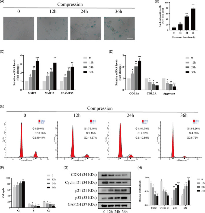
Inappropriate or excessive compression applied to intervertebral disc (IVD) contributes substantially to IVD degeneration. The actomyosin system plays a leading role in responding to mechanical stimuli. In the present study, we investigated the roles of myosin II isoforms in the compression stress-induced senescence of nucleus pulposus (NP) cells.
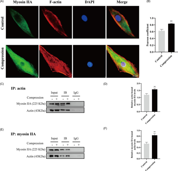
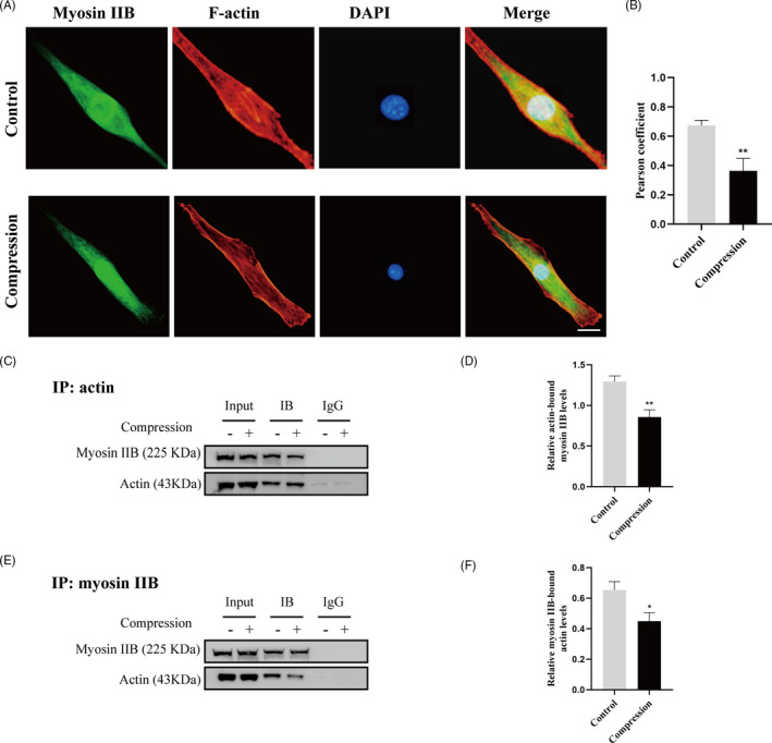
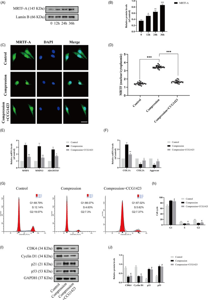
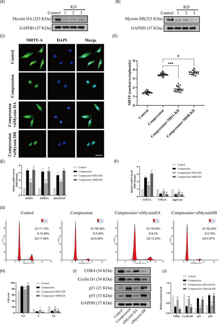
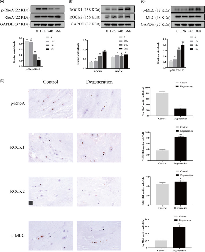
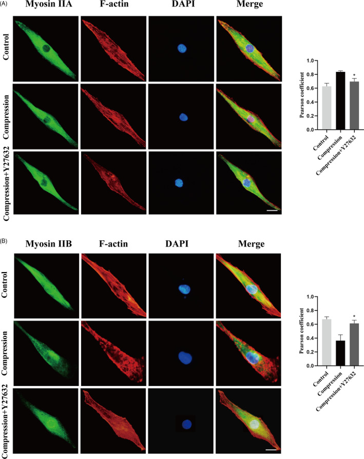
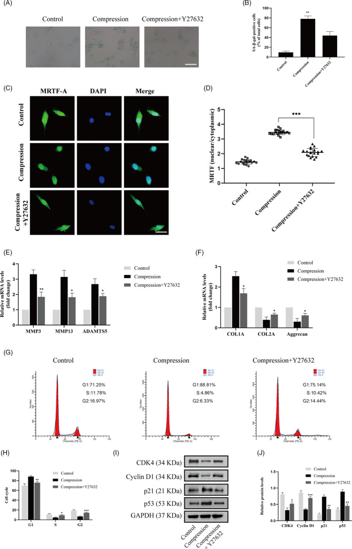
Nucleus pulposus cells were exposed to 1.0 MPa compression for 0, 12, 24 or 36 hours. Immunofluorescence and co-immunoprecipitation analysis were used to measure the interaction of myosin IIA and IIB with actin. Western blot analysis and immunofluorescence staining were used to detect nuclear expression and nuclear localization of MRTF-A. In addition, the expression levels of p-RhoA/RhoA, ROCK1/2 and p-MLC/MLC were measured in human NP cells under compression stress and in degenerative IVD tissues.
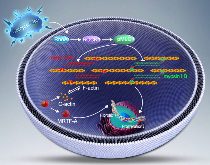
Compression stress increased the interaction of myosin IIA and actin, while the interaction of myosin IIB and actin was reduced. The actomyosin cytoskeleton remodelling was involved in the compression stress-induced fibrotic phenotype mediated by MRTF-A nuclear translocation and inhibition of proliferation in NP cells. Furthermore, RhoA/ROCK1 pathway activation mediated compression stress-induced human NP cells senescence by regulating the interaction of myosin IIA and IIB with actin.
We for the first time investigated the regulation of actomyosin cytoskeleton in human NP cells under compression stress. It provided new insights into the development of therapy for effectively inhibiting IVD degeneration.
施加于椎间盘(IVD)的不当或过度压缩会极大地促进 IVD 退化。肌球蛋白 II 系统在响应机械刺激方面起着主导作用。在本研究中,我们研究了肌球蛋白 II 同工型在椎间盘核(NP)细胞压缩应激诱导衰老中的作用。
NP 细胞暴露于 1.0 MPa 压缩下 0、12、24 或 36 小时。免疫荧光和共免疫沉淀分析用于测量肌球蛋白 IIA 和 IIB 与肌动蛋白的相互作用。Western blot 分析和免疫荧光染色用于检测 MRTF-A 的核表达和核定位。此外,在受压的人 NP 细胞和退行性 IVD 组织中测量了 p-RhoA/RhoA、ROCK1/2 和 p-MLC/MLC 的表达水平。
压缩应激增加了肌球蛋白 IIA 与肌动蛋白的相互作用,而肌球蛋白 IIB 与肌动蛋白的相互作用减少。肌动球蛋白细胞骨架重塑参与了由 MRTF-A 核转位介导的压缩应激诱导的 NP 细胞纤维化表型,并抑制了 NP 细胞的增殖。此外,RhoA/ROCK1 通路的激活通过调节肌球蛋白 IIA 和 IIB 与肌动蛋白的相互作用,介导了压缩应激诱导的人 NP 细胞衰老。
我们首次研究了人 NP 细胞在压缩应激下肌动球蛋白细胞骨架的调节。这为开发有效抑制 IVD 退化的治疗方法提供了新的见解。