• 文献检索
  • 文档翻译
  • 深度研究
  • 学术资讯
  • Suppr Zotero 插件Zotero 插件
  • 邀请有礼
  • 套餐&价格
  • 历史记录
应用&插件
Suppr Zotero 插件Zotero 插件浏览器插件Mac 客户端Windows 客户端微信小程序
定价
高级版会员购买积分包购买API积分包
服务
文献检索文档翻译深度研究API 文档MCP 服务
关于我们
关于 Suppr公司介绍联系我们用户协议隐私条款
关注我们

Suppr 超能文献

核心技术专利:CN118964589B侵权必究
粤ICP备2023148730 号-1Suppr @ 2026

文献检索

告别复杂PubMed语法,用中文像聊天一样搜索,搜遍4000万医学文献。AI智能推荐,让科研检索更轻松。

立即免费搜索

文件翻译

保留排版,准确专业,支持PDF/Word/PPT等文件格式,支持 12+语言互译。

免费翻译文档

深度研究

AI帮你快速写综述,25分钟生成高质量综述,智能提取关键信息,辅助科研写作。

立即免费体验

利用扫描电子辅助介电显微镜观察 PM2.5 进入哺乳动物细胞的纳米级结构。

Nanoscale observation of PM2.5 incorporated into mammalian cells using scanning electron-assisted dielectric microscope.

机构信息

Health and Medical Research Institute, National Institute of Advanced Industrial Science and Technology (AIST), Central 6, Higashi 1-1-1, Tsukuba, Ibaraki, 305-8566, Japan.

Department of Periodontology, Osaka University Graduate School of Dentistry, 1-8 Yamada-oka, Suita, Osaka, 565-0851, Japan.

出版信息

Sci Rep. 2021 Jan 8;11(1):228. doi: 10.1038/s41598-020-80546-0.

DOI:10.1038/s41598-020-80546-0
PMID:33420286
原文链接:https://pmc.ncbi.nlm.nih.gov/articles/PMC7794539/
Abstract

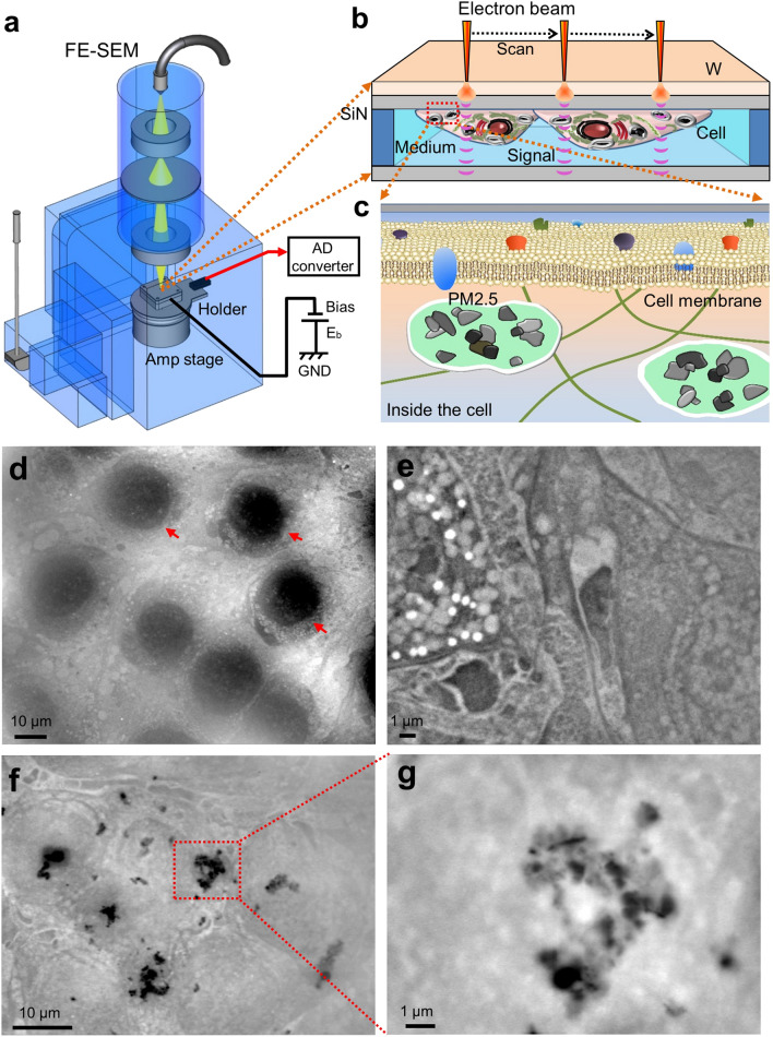
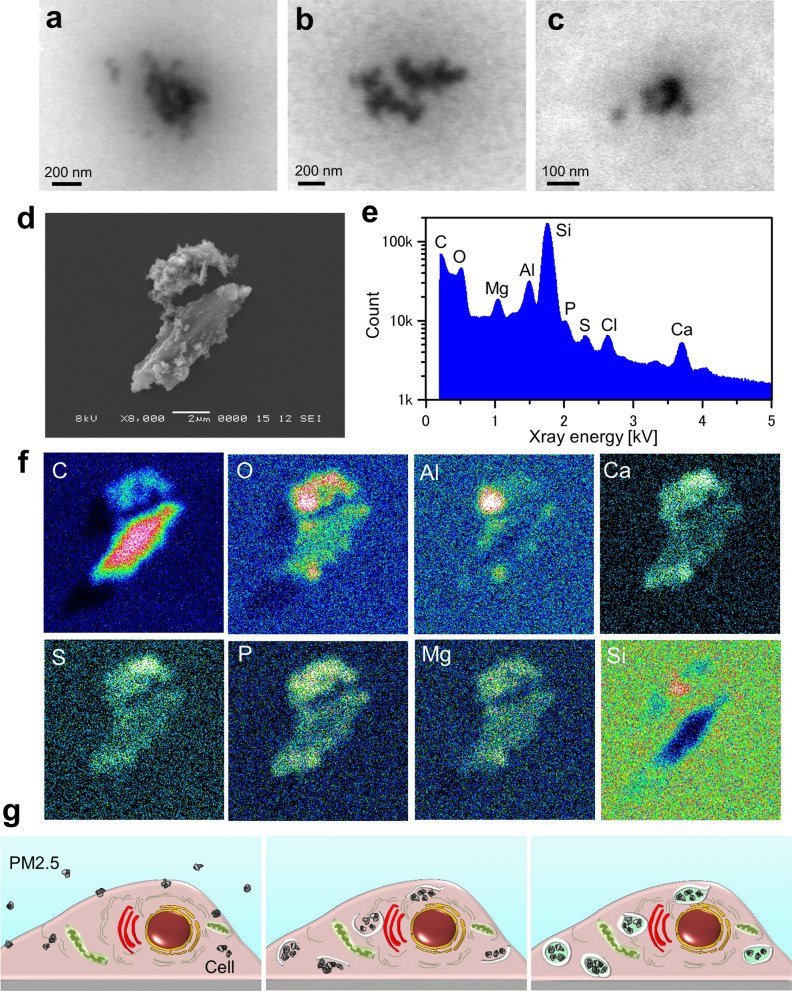
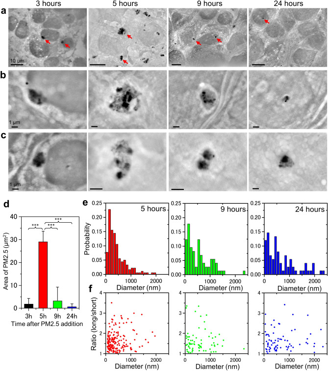
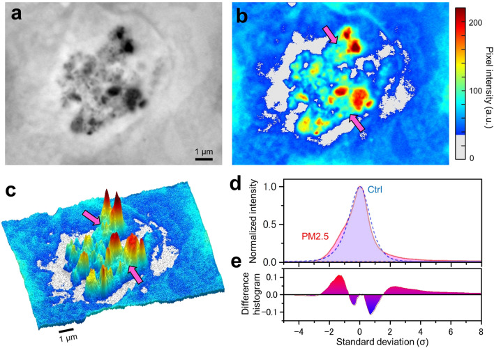
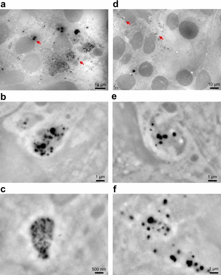
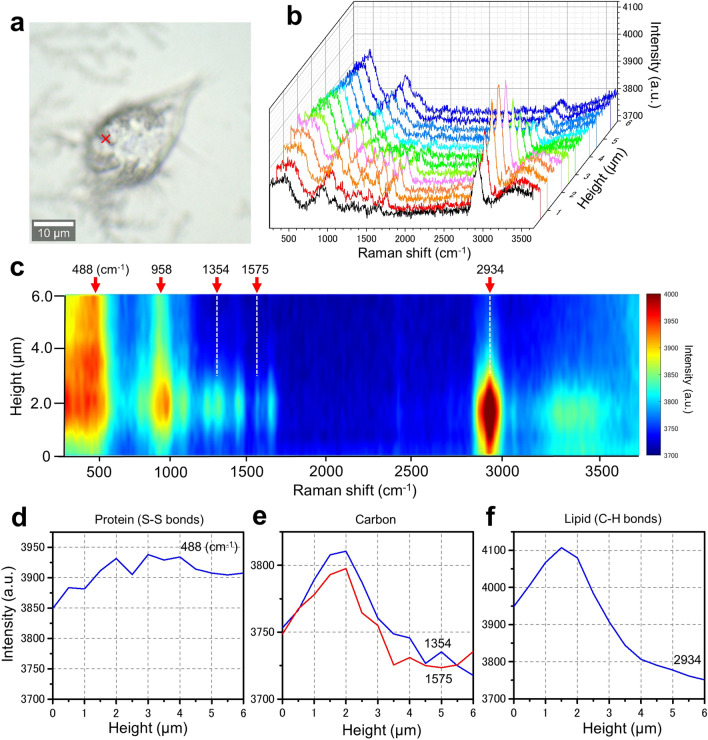
PM2.5 has been correlated with risk factors for various diseases and infections. It promotes tissue injury by direct effects of particle components. However, effects of PM2.5 on cells have not been fully investigated. Recently, we developed a novel imaging technology, scanning electron-assisted dielectric-impedance microscopy (SE-ADM), which enables observation of various biological specimens in aqueous solution. In this study, we successfully observed PM2.5 incorporated into living mammalian cells in culture media. Our system directly revealed the process of PM2.5 aggregation in the cells at a nanometre resolution. Further, we found that the PM2.5 aggregates in the intact cells were surrounded by intracellular membrane-like structures of low-density in the SE-ADM images. Moreover, the PM2.5 aggregates were shown by confocal Raman microscopy to be located inside the cells rather than on the cell surface. We expect our method to be applicable to the observation of various nanoparticles inside cells in culture media.

摘要

PM2.5 与各种疾病和感染的风险因素有关。它通过颗粒成分的直接作用促进组织损伤。然而,PM2.5 对细胞的影响尚未得到充分研究。最近,我们开发了一种新型成像技术,扫描电子辅助介电阻抗显微镜(SE-ADM),它可以观察水相中的各种生物样本。在这项研究中,我们成功地观察到 PM2.5 掺入培养介质中的活哺乳动物细胞。我们的系统以纳米分辨率直接揭示了 PM2.5 在细胞内聚集的过程。此外,我们发现,在 SE-ADM 图像中,完整细胞内的 PM2.5 聚集体被低密度的细胞内类似膜结构包围。此外,共聚焦拉曼显微镜显示 PM2.5 聚集体位于细胞内,而不是细胞表面。我们期望我们的方法适用于观察培养介质中细胞内各种纳米颗粒。

https://cdn.ncbi.nlm.nih.gov/pmc/blobs/5de0/7794539/2601e3285681/41598_2020_80546_Fig6_HTML.jpg
https://cdn.ncbi.nlm.nih.gov/pmc/blobs/5de0/7794539/5e6442428f76/41598_2020_80546_Fig1_HTML.jpg
https://cdn.ncbi.nlm.nih.gov/pmc/blobs/5de0/7794539/f8f0912b6226/41598_2020_80546_Fig2_HTML.jpg
https://cdn.ncbi.nlm.nih.gov/pmc/blobs/5de0/7794539/222d00679f79/41598_2020_80546_Fig3_HTML.jpg
https://cdn.ncbi.nlm.nih.gov/pmc/blobs/5de0/7794539/e0e4935f8bc8/41598_2020_80546_Fig4_HTML.jpg
https://cdn.ncbi.nlm.nih.gov/pmc/blobs/5de0/7794539/dfa51526795b/41598_2020_80546_Fig5_HTML.jpg
https://cdn.ncbi.nlm.nih.gov/pmc/blobs/5de0/7794539/2601e3285681/41598_2020_80546_Fig6_HTML.jpg

相似文献

1
Nanoscale observation of PM2.5 incorporated into mammalian cells using scanning electron-assisted dielectric microscope.利用扫描电子辅助介电显微镜观察 PM2.5 进入哺乳动物细胞的纳米级结构。
Sci Rep. 2021 Jan 8;11(1):228. doi: 10.1038/s41598-020-80546-0.
2
Development of General-purpose Dielectric Constant Imaging Unit for SEM and Direct Observation of Samples in Aqueous Solution.用于扫描电子显微镜的通用介电常数成像单元的开发及水溶液中样品的直接观察。
Microsc Microanal. 2023 Jun 9;29(3):1037-1046. doi: 10.1093/micmic/ozad030.
3
High-resolution imaging of living mammalian cells bound by nanobeads-connected antibodies in a medium using scanning electron-assisted dielectric microscopy.利用扫描电子辅助介电显微镜对结合有纳米珠连接抗体的活哺乳动物细胞在介质中的高分辨率成像。
Sci Rep. 2017 Feb 23;7:43025. doi: 10.1038/srep43025.
4
Nanoscale imaging of untreated mammalian cells in a medium with low radiation damage using scanning electron-assisted dielectric microscopy.使用扫描电子辅助介电显微镜对低辐射损伤介质中的未经处理的哺乳动物细胞进行纳米级成像。
Sci Rep. 2016 Jul 4;6:29169. doi: 10.1038/srep29169.
5
Nanoscale imaging of the adhesion core including integrin β1 on intact living cells using scanning electron-assisted dielectric-impedance microscopy.使用扫描电子辅助介电阻抗显微镜对完整活细胞上的整合素β1 等粘附核心进行纳米级成像。
PLoS One. 2018 Sep 20;13(9):e0204133. doi: 10.1371/journal.pone.0204133. eCollection 2018.
6
Nanoscale observation of the natural structure of milk-fat globules and casein micelles in the liquid condition using a scanning electron assisted dielectric microscopy.使用扫描电子辅助介电显微镜对液态条件下乳脂肪球和酪蛋白胶束的天然结构进行纳米级观察。
Biochem Biophys Res Commun. 2017 Sep 30;491(4):1021-1025. doi: 10.1016/j.bbrc.2017.08.004. Epub 2017 Aug 2.
7
Nano-Microscopy of Therapeutic Antibody Aggregates in Solution.溶液中治疗性抗体聚集体的纳米显微镜研究
Methods Mol Biol. 2022;2313:219-239. doi: 10.1007/978-1-0716-1450-1_13.
8
Nanoscale characterization of PM2.5 airborne pollutants reveals high adhesiveness and aggregation capability of soot particles.细颗粒物(PM2.5)空气污染物的纳米级表征揭示了烟尘颗粒具有高粘附性和聚集能力。
Sci Rep. 2015 Jul 16;5:11232. doi: 10.1038/srep11232.
9
Scanning Electron-Assisted Dielectric Microscopy Reveals Autophagosome Formation by LC3 and ATG12 in Cultured Mammalian Cells.扫描电子显微镜辅助介电泳显微镜观察到培养的哺乳动物细胞中 LC3 和 ATG12 诱导自噬体的形成。
Int J Mol Sci. 2021 Feb 12;22(4):1834. doi: 10.3390/ijms22041834.
10
Interaction of PM2.5 airborne particulates with ZnO and TiO nanoparticles and their effect on bacteria.空气中细颗粒物PM2.5与氧化锌和二氧化钛纳米颗粒的相互作用及其对细菌的影响。
Environ Monit Assess. 2017 Dec 21;190(1):34. doi: 10.1007/s10661-017-6408-2.

引用本文的文献

1
PM, component cause of severe metabolically abnormal obesity: An in silico, observational and analytical study.PM,严重代谢异常肥胖的构成原因:一项计算机模拟、观察性及分析性研究。
Heliyon. 2024 Apr 3;10(7):e28936. doi: 10.1016/j.heliyon.2024.e28936. eCollection 2024 Apr 15.
2
Structural analysis of melanosomes in living mammalian cells using scanning electron-assisted dielectric microscopy with deep neural network.使用扫描电子辅助介电显微镜和深度神经网络对活的哺乳动物细胞中的黑素小体进行结构分析。
Comput Struct Biotechnol J. 2022 Dec 18;21:506-518. doi: 10.1016/j.csbj.2022.12.027. eCollection 2023.
3
Raman scattering enhancement of dielectric microspheres on silicon nitride film.

本文引用的文献

1
Environmental determinants of cardiovascular disease: lessons learned from air pollution.心血管疾病的环境决定因素:空气污染带来的启示。
Nat Rev Cardiol. 2020 Oct;17(10):656-672. doi: 10.1038/s41569-020-0371-2. Epub 2020 May 7.
2
Osteoblastic lysosome plays a central role in mineralization.成骨细胞溶酶体在矿化中起核心作用。
Sci Adv. 2019 Jul 3;5(7):eaax0672. doi: 10.1126/sciadv.aax0672. eCollection 2019 Jul.
3
Monitoring glycosylation metabolism in brain and breast cancer by Raman imaging.通过拉曼成像监测大脑和乳腺癌中的糖基化代谢。
氮化硅薄膜上介质微球的喇曼散射增强。
Sci Rep. 2022 Mar 29;12(1):5346. doi: 10.1038/s41598-022-09315-5.
4
Scanning Electron-Assisted Dielectric Microscopy Reveals Autophagosome Formation by LC3 and ATG12 in Cultured Mammalian Cells.扫描电子显微镜辅助介电泳显微镜观察到培养的哺乳动物细胞中 LC3 和 ATG12 诱导自噬体的形成。
Int J Mol Sci. 2021 Feb 12;22(4):1834. doi: 10.3390/ijms22041834.
Sci Rep. 2019 Jan 17;9(1):166. doi: 10.1038/s41598-018-36622-7.
4
Nanoscale imaging of the adhesion core including integrin β1 on intact living cells using scanning electron-assisted dielectric-impedance microscopy.使用扫描电子辅助介电阻抗显微镜对完整活细胞上的整合素β1 等粘附核心进行纳米级成像。
PLoS One. 2018 Sep 20;13(9):e0204133. doi: 10.1371/journal.pone.0204133. eCollection 2018.
5
COX-2 mediates PM2.5-induced apoptosis and inflammation in vascular endothelial cells.环氧化酶-2介导细颗粒物2.5诱导的血管内皮细胞凋亡和炎症反应。
Am J Transl Res. 2017 Sep 15;9(9):3967-3976. eCollection 2017.
6
Nanoscale observation of the natural structure of milk-fat globules and casein micelles in the liquid condition using a scanning electron assisted dielectric microscopy.使用扫描电子辅助介电显微镜对液态条件下乳脂肪球和酪蛋白胶束的天然结构进行纳米级观察。
Biochem Biophys Res Commun. 2017 Sep 30;491(4):1021-1025. doi: 10.1016/j.bbrc.2017.08.004. Epub 2017 Aug 2.
7
Stress fibers, autophagy and necrosis by persistent exposure to PM2.5 from biomass combustion.长期暴露于生物质燃烧产生的细颗粒物(PM2.5)导致的应力纤维、自噬和坏死
PLoS One. 2017 Jul 3;12(7):e0180291. doi: 10.1371/journal.pone.0180291. eCollection 2017.
8
Airborne particulate matter (PM2.5) triggers autophagy in human corneal epithelial cell line.空气中的颗粒物(PM2.5)可触发人角膜上皮细胞系中的自噬。
Environ Pollut. 2017 Aug;227:314-322. doi: 10.1016/j.envpol.2017.04.078. Epub 2017 May 4.
9
Exposure scenario: Another important factor determining the toxic effects of PM2.5 and possible mechanisms involved.暴露情景:另一个决定 PM2.5 毒性效应和可能涉及的机制的重要因素。
Environ Pollut. 2017 Jul;226:412-425. doi: 10.1016/j.envpol.2017.04.010. Epub 2017 Apr 25.
10
Physicochemical characterization of ambient PM in Tehran air and its potential cytotoxicity in human lung epithelial cells (A549).德黑兰空气环境 PM 的理化特性及其对人肺上皮细胞(A549)的潜在细胞毒性。
Sci Total Environ. 2017 Sep 1;593-594:182-190. doi: 10.1016/j.scitotenv.2017.03.150. Epub 2017 Mar 23.