• 文献检索
  • 文档翻译
  • 深度研究
  • 学术资讯
  • Suppr Zotero 插件Zotero 插件
  • 邀请有礼
  • 套餐&价格
  • 历史记录
应用&插件
Suppr Zotero 插件Zotero 插件浏览器插件Mac 客户端Windows 客户端微信小程序
定价
高级版会员购买积分包购买API积分包
服务
文献检索文档翻译深度研究API 文档MCP 服务
关于我们
关于 Suppr公司介绍联系我们用户协议隐私条款
关注我们

Suppr 超能文献

核心技术专利:CN118964589B侵权必究
粤ICP备2023148730 号-1Suppr @ 2026

文献检索

告别复杂PubMed语法,用中文像聊天一样搜索,搜遍4000万医学文献。AI智能推荐,让科研检索更轻松。

立即免费搜索

文件翻译

保留排版,准确专业,支持PDF/Word/PPT等文件格式,支持 12+语言互译。

免费翻译文档

深度研究

AI帮你快速写综述,25分钟生成高质量综述,智能提取关键信息,辅助科研写作。

立即免费体验

多发性硬化症中核间性眼肌麻痹的临床-放射学悖论探讨模型

A model for interrogating the clinico-radiological paradox in multiple sclerosis: Internuclear ophthalmoplegia.

机构信息

Department of Neurology, MS Center and Neuro-ophthalmology Expertise Center, Amsterdam Neuroscience, Amsterdam UMC, Vrije Universiteit Amsterdam, Amsterdam, The Netherlands.

Department of Ophthalmology, Neuro-ophthalmology Expertise Center, Amsterdam Neuroscience, Amsterdam UMC, Vrije Universiteit Amsterdam, Amsterdam, The Netherlands.

出版信息

Eur J Neurol. 2021 May;28(5):1617-1626. doi: 10.1111/ene.14723. Epub 2021 Feb 8.

DOI:10.1111/ene.14723
PMID:33426786
原文链接:https://pmc.ncbi.nlm.nih.gov/articles/PMC8248033/
Abstract

BACKGROUND AND PURPOSE

The clinico-radiological paradox in multiple sclerosis (MS) is well recognized, relevant and yet poorly understood. The suitability of an in vivo model for the clinico-radiological paradox was tested, using internuclear ophthalmoplegia (INO) and the medial longitudinal fasciculus (MLF).

METHODS

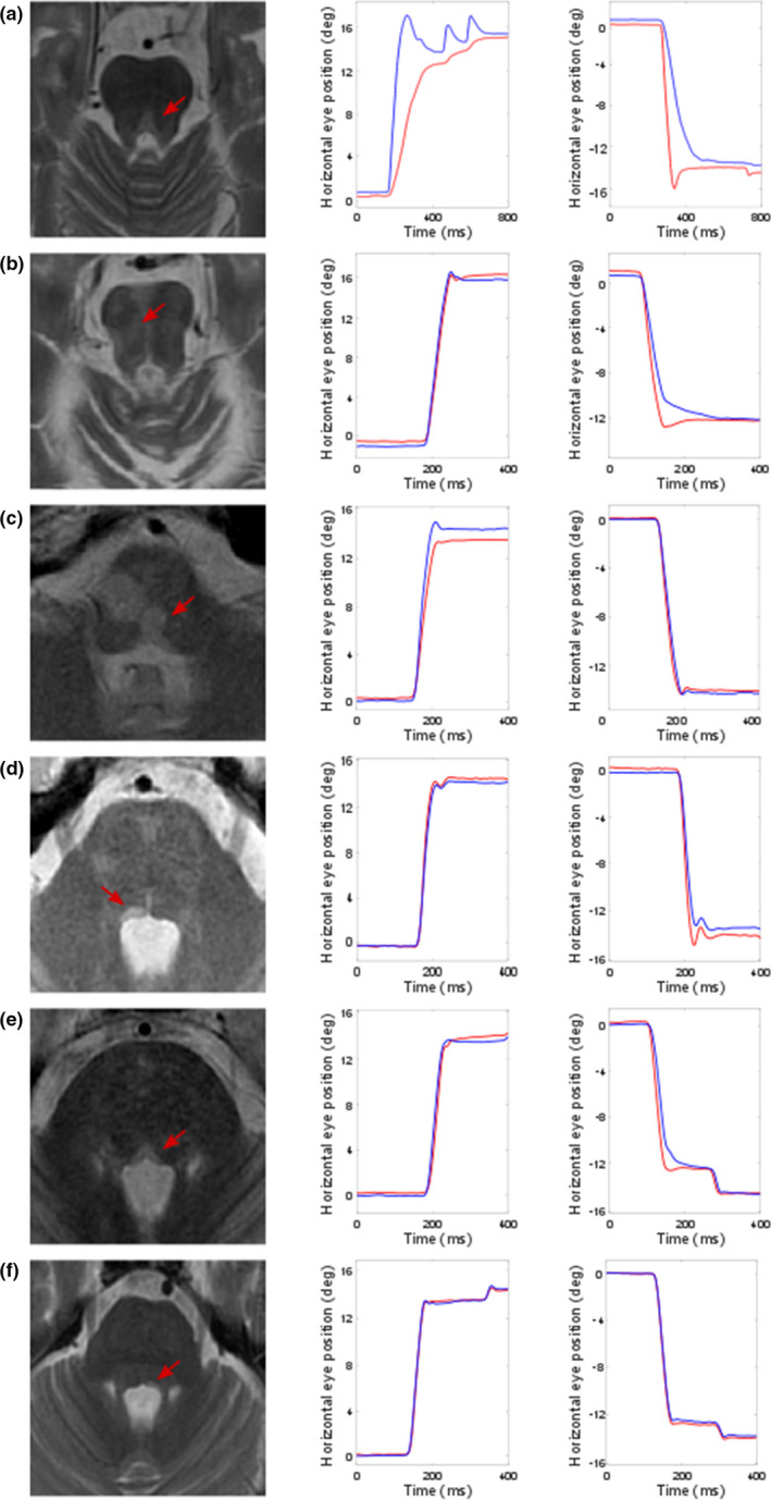
In this cross-sectional study lesions of the MLF were rated by an experienced MS neuroradiologist blinded to all other information. The presence of an INO was objectively determined by a validated infrared oculography protocol (DEMoNS). Clinical information, including the National Eye Institute Visual Function Questionnaire, was obtained.

RESULTS

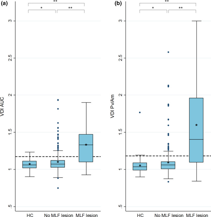
This study included 202 patients with MS. The clinico-radiological paradox occurred in 50 patients (25%). This consisted of 45 patients having an INO without an MLF lesion and five patients with an MLF lesion but without an INO. The visual function overall score was related to the presence of an INO (p = 0.016), but not to MLF lesions seen on magnetic resonance imaging (MRI) (p = 0.207). A consensus list of potential causes for the clinico-radiological paradox was compiled and the MRI images were deposited in a repository.

CONCLUSION

This study provides an objective and quantitative model to investigate the clinico-radiological paradox. Our data suggest that pathology of the MLF is more frequently detected and more clinically relevant by infrared oculography than by MLF lesion rating on MRI.

摘要

背景与目的

多发性硬化症(MS)的临床-影像学悖论是公认的、相关的,但理解不足。本研究通过核间性眼肌麻痹(INO)和内侧纵束(MLF)来测试体内模型对临床-影像学悖论的适用性。

方法

本横断面研究由一位经验丰富的 MS 神经放射科医生对 MLF 病变进行评分,该医生对所有其他信息均不知情。通过经过验证的红外眼动图协议(DEMoNS)客观确定 INO 的存在。获取临床信息,包括美国国立眼科研究所视觉功能问卷。

结果

本研究纳入了 202 例 MS 患者。50 例(25%)患者存在临床-影像学悖论。具体表现为 45 例患者存在 INO 但无 MLF 病变,5 例患者存在 MLF 病变但无 INO。总体视觉功能评分与 INO 的存在相关(p=0.016),但与 MRI 上观察到的 MLF 病变无关(p=0.207)。我们编制了一份潜在临床-影像学悖论原因的共识清单,并将 MRI 图像存入存储库。

结论

本研究提供了一种客观、定量的方法来研究临床-影像学悖论。我们的数据表明,与 MRI 上 MLF 病变评分相比,红外眼动图更能经常检测到 MLF 的病理学改变,且更具有临床相关性。

https://cdn.ncbi.nlm.nih.gov/pmc/blobs/6257/8248033/2ff98381192c/ENE-28-1617-g001.jpg
https://cdn.ncbi.nlm.nih.gov/pmc/blobs/6257/8248033/a6efddf858c8/ENE-28-1617-g003.jpg
https://cdn.ncbi.nlm.nih.gov/pmc/blobs/6257/8248033/2ff98381192c/ENE-28-1617-g001.jpg
https://cdn.ncbi.nlm.nih.gov/pmc/blobs/6257/8248033/a6efddf858c8/ENE-28-1617-g003.jpg
https://cdn.ncbi.nlm.nih.gov/pmc/blobs/6257/8248033/2ff98381192c/ENE-28-1617-g001.jpg

相似文献

1
A model for interrogating the clinico-radiological paradox in multiple sclerosis: Internuclear ophthalmoplegia.多发性硬化症中核间性眼肌麻痹的临床-放射学悖论探讨模型
Eur J Neurol. 2021 May;28(5):1617-1626. doi: 10.1111/ene.14723. Epub 2021 Feb 8.
2
Lesion follows function: video-oculography compared with MRI to diagnose internuclear ophthalmoplegia in patients with multiple sclerosis.病灶随功能而变化:视频眼震图与 MRI 对比诊断多发性硬化患者的核间性眼肌麻痹。
J Neurol. 2023 Feb;270(2):917-924. doi: 10.1007/s00415-022-11428-w. Epub 2022 Oct 31.
3
Sensitivity of Magnetic Resonance Imaging of the Medial Longitudinal Fasciculus in Internuclear Ophthalmoplegia.内侧纵束核间性眼肌麻痹的磁共振成像敏感性。
J Neuroophthalmol. 2024 Mar 1;44(1):107-111. doi: 10.1097/WNO.0000000000001783. Epub 2023 Jan 4.
4
Visualisation of the medial longitudinal fasciculus using fibre tractography in multiple sclerosis patients with internuclear ophthalmoplegia.在患有核间性眼肌麻痹的多发性硬化症患者中使用纤维束成像技术对内侧纵束进行可视化。
Ir J Med Sci. 2016 May;185(2):393-402. doi: 10.1007/s11845-016-1405-y. Epub 2016 Jan 19.
5
Vestibulo-ocular reflex deficits with medial longitudinal fasciculus lesions.前庭眼反射缺陷伴内侧纵束病变。
J Neurol. 2017 Oct;264(10):2119-2129. doi: 10.1007/s00415-017-8607-8. Epub 2017 Sep 6.
6
MRI topography of lesions related to internuclear ophthalmoplegia in patients with multiple sclerosis or ischemic stroke.MRI topography of lesions related to internuclear ophthalmoplegia in patients with multiple sclerosis or ischemic stroke.
J Neuroimaging. 2021 May;31(3):471-474. doi: 10.1111/jon.12847. Epub 2021 Apr 1.
7
[Internuclear ophthalmoplegia in course of multiple sclerosis and head trauma. Presentation of 2 cases].[多发性硬化症和头部外伤病程中的核间性眼肌麻痹。2例报告]
Klin Oczna. 2009;111(4-6):156-9.
8
MRI characteristics of the MLF in MS patients with chronic internuclear ophthalmoparesis.患有慢性核间性眼肌麻痹的多发性硬化症患者中内侧纵束的磁共振成像特征。
Neurology. 2001 Sep 11;57(5):762-8. doi: 10.1212/wnl.57.5.762.
9
Axonal conduction in multiple sclerosis: A combined magnetic resonance imaging and electrophysiological study of the medial longitudinal fasciculus.多发性硬化症中的轴突传导:内侧纵束的磁共振成像与电生理联合研究
Mult Scler. 2015 Jun;21(7):905-15. doi: 10.1177/1352458514556301. Epub 2014 Nov 12.
10
Diagnosing and quantifying a common deficit in multiple sclerosis: Internuclear ophthalmoplegia.诊断和量化多发性硬化症的常见缺陷:眼肌神经核间麻痹。
Neurology. 2019 May 14;92(20):e2299-e2308. doi: 10.1212/WNL.0000000000007499. Epub 2019 Apr 19.

引用本文的文献

1
Ultra-High Contrast (UHC) MRI of the Brain, Spinal Cord and Optic Nerves in Multiple Sclerosis Using Directly Acquired and Synthetic Bipolar Filter (BLAIR) Images.使用直接采集和合成双极滤波器(BLAIR)图像对多发性硬化症患者的脑、脊髓和视神经进行超高对比度(UHC)磁共振成像
Diagnostics (Basel). 2025 Jan 30;15(3):329. doi: 10.3390/diagnostics15030329.
2
The virtual multiple sclerosis patient.虚拟多发性硬化症患者。
iScience. 2024 May 24;27(7):110101. doi: 10.1016/j.isci.2024.110101. eCollection 2024 Jul 19.
3
Clinical-Radiological Mismatch in Multiple Sclerosis Patients during Acute Relapse: Discrepancy between Clinical Symptoms and Active, Topographically Fitting MRI Lesions.

本文引用的文献

1
The neuroanatomical-functional paradox in spinal cord injury.脊髓损伤的神经解剖-功能悖论。
Nat Rev Neurol. 2021 Jan;17(1):53-62. doi: 10.1038/s41582-020-00436-x. Epub 2020 Dec 11.
2
Using Acute Optic Neuritis Trials to Assess Neuroprotective and Remyelinating Therapies in Multiple Sclerosis.利用急性视神经炎试验评估多发性硬化症的神经保护和髓鞘修复疗法。
JAMA Neurol. 2020 Feb 1;77(2):234-244. doi: 10.1001/jamaneurol.2019.3283.
3
Diagnosing and quantifying a common deficit in multiple sclerosis: Internuclear ophthalmoplegia.
多发性硬化症患者急性复发期的临床-放射学不匹配:临床症状与活跃的、地形学上匹配的MRI病变之间的差异
J Clin Med. 2023 Jan 17;12(3):739. doi: 10.3390/jcm12030739.
诊断和量化多发性硬化症的常见缺陷:眼肌神经核间麻痹。
Neurology. 2019 May 14;92(20):e2299-e2308. doi: 10.1212/WNL.0000000000007499. Epub 2019 Apr 19.
4
Quantification of Visual Fixation in Multiple Sclerosis.多发性硬化症的视觉固视定量。
Invest Ophthalmol Vis Sci. 2019 Apr 1;60(5):1372-1383. doi: 10.1167/iovs.18-26096.
5
Treatment of internuclear ophthalmoparesis in multiple sclerosis with fampridine: A randomized double-blind, placebo-controlled cross-over trial.多发性硬化症核间性眼肌麻痹的治疗:一项随机、双盲、安慰剂对照交叉试验。
CNS Neurosci Ther. 2019 Jun;25(6):697-703. doi: 10.1111/cns.13096. Epub 2019 Feb 12.
6
A standardized protocol for quantification of saccadic eye movements: DEMoNS.用于扫视眼动定量的标准化方案:DEMoNS。
PLoS One. 2018 Jul 16;13(7):e0200695. doi: 10.1371/journal.pone.0200695. eCollection 2018.
7
Assessing Biological and Methodological Aspects of Brain Volume Loss in Multiple Sclerosis.评估多发性硬化症中脑容量损失的生物学和方法学方面。
JAMA Neurol. 2018 Oct 1;75(10):1246-1255. doi: 10.1001/jamaneurol.2018.1596.
8
Remodeling Functional Connectivity in Multiple Sclerosis: A Challenging Therapeutic Approach.重塑多发性硬化症中的功能连接:一种具有挑战性的治疗方法。
Front Neurosci. 2017 Dec 13;11:710. doi: 10.3389/fnins.2017.00710. eCollection 2017.
9
Retinal atrophy in relation to visual functioning and vision-related quality of life in patients with multiple sclerosis.多发性硬化症患者的视网膜萎缩与视觉功能和与视觉相关的生活质量有关。
Mult Scler. 2018 May;24(6):767-776. doi: 10.1177/1352458517708463. Epub 2017 May 17.
10
The clinico-radiological paradox of cognitive function and MRI burden of white matter lesions in people with multiple sclerosis: A systematic review and meta-analysis.多发性硬化症患者认知功能与脑白质病变MRI负荷的临床-放射学悖论:一项系统评价和荟萃分析
PLoS One. 2017 May 15;12(5):e0177727. doi: 10.1371/journal.pone.0177727. eCollection 2017.