Shapira Nadav, Mei Kai, Noël Peter B
Department of Radiology, University of Pennsylvania, Philadelphia, USA.
Department of Diagnostic and Interventional Radiology, Technical University of Munich, Munich, Germany.
J Appl Clin Med Phys. 2021 Mar;22(3):16-26. doi: 10.1002/acm2.13161. Epub 2021 Jan 10.
Spectral computed tomography (spectral CT) provides access to clinically relevant measures of endogenous and exogenous materials in patients. For pediatric patients, current spectral CT applications include lesion characterization, quantitative vascular imaging, assessments of tumor response to treatment, and more.
The aim of this study is a comprehensive investigation of the accuracy and stability of spectral quantifications from a spectral detector-based CT system with respect to different patient sizes and radiation dose levels relevant for the pediatric population.
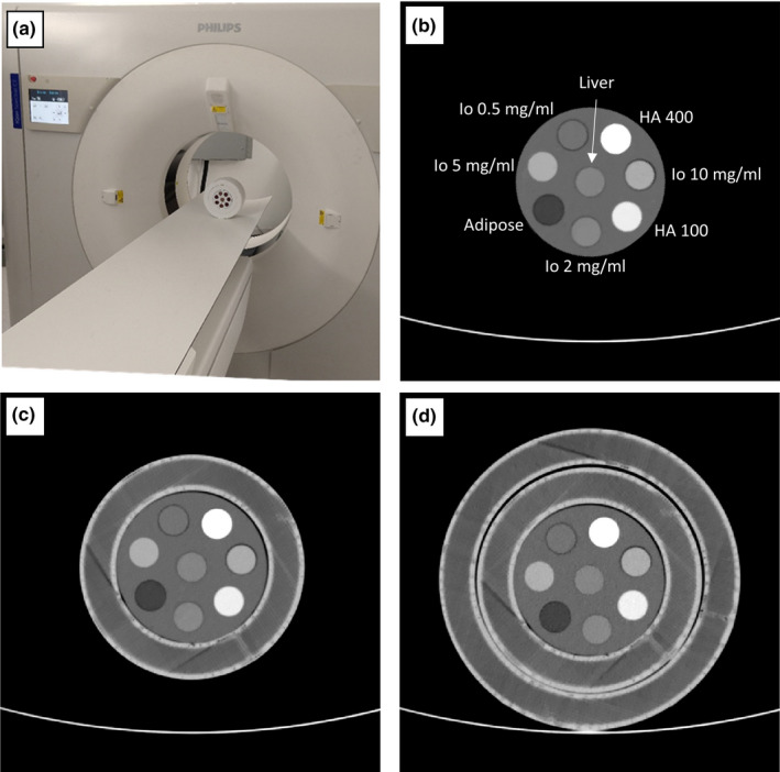
A spectral CT phantom with tissue-mimicking materials and iodine concentrations relevant for pediatric imaging was scanned on a spectral detector CT system using a standard pediatric abdominal protocol at 100%, 67%, 33% and 10% of the nominal radiation dose level. Different pediatric patient sizes were simulated using supplemental 3D-printed extension rings. Virtual mono-energetic, iodine density, effective atomic number, and electron density results were analyzed for stability with respect to radiation dose and patient size.
Compared to conventional CT imaging, a pronounced improvement in the stability of attenuation measurements across patient size was observed when using virtual mono-energetic images. Iodine densities were within 0.1 mg/ml, effective atomic numbers were within 0.26 atomic numbers and electron density quantifications were within ±1.0% of their respective nominal values. Relative to the nominal dose clinical protocol, differences in attenuation of all tissue-mimicking materials were maintained below 1.6 HU for a 33% dose reduction, below 2.7 HU for a 67% dose reduction and below 3.7 HU for a 90% dose reduction, for all virtual mono-energetic energies equal to or greater than 50 keV. Iodine, and effective atomic number quantifications were stable to within 0.1 mg/ml and 0.06 atomic numbers, respectively, across all measured dose levels.
Spectral CT provides accurate and stable material quantification with respect to radiation dose reduction (up to 90%) and differing pediatric patient size. The observed consistency is an important step towards quantitative pediatric imaging at low radiation exposure levels.
光谱计算机断层扫描(光谱CT)能够获取患者体内内源性和外源性物质的临床相关测量值。对于儿科患者,当前光谱CT的应用包括病变特征描述、定量血管成像、评估肿瘤对治疗的反应等。
本研究旨在全面调查基于光谱探测器的CT系统在与儿科人群相关的不同患者体型和辐射剂量水平下,光谱定量的准确性和稳定性。
使用标准儿科腹部扫描方案,在光谱探测器CT系统上,以标称辐射剂量水平的100%、67%、33%和10%对含有与儿科成像相关的组织模拟材料和碘浓度的光谱CT体模进行扫描。使用补充的3D打印延伸环模拟不同的儿科患者体型。分析虚拟单能、碘密度、有效原子序数和电子密度结果在辐射剂量和患者体型方面的稳定性。
与传统CT成像相比,使用虚拟单能图像时,观察到跨患者体型的衰减测量稳定性有显著提高。碘密度在0.1mg/ml以内,有效原子序数在0.26个原子序数以内,电子密度定量在其各自标称值的±1.0%以内。相对于标称剂量临床方案,对于所有等于或大于50keV的虚拟单能能量,当剂量降低33%时,所有组织模拟材料的衰减差异保持在1.6HU以下;当剂量降低67%时,低于2.7HU;当剂量降低90%时,低于3.7HU。在所有测量的剂量水平上,碘和有效原子序数定量分别稳定在0.1mg/ml和0.06个原子序数以内。
光谱CT在降低辐射剂量(高达90%)和不同儿科患者体型的情况下,提供了准确且稳定的物质定量。观察到的一致性是朝着低辐射暴露水平下的儿科定量成像迈出的重要一步。