Department of Neurosurgery, University of Tuebingen, Hoppe-Seyler-Str. 3, 72076, Tuebingen, Germany.
Department of Neuroradiology, University of Tuebingen, Hoppe-Seyler-Str. 3, 72076, Tuebingen, Germany.
Neurosurg Rev. 2021 Oct;44(5):2947-2956. doi: 10.1007/s10143-020-01468-z. Epub 2021 Jan 11.
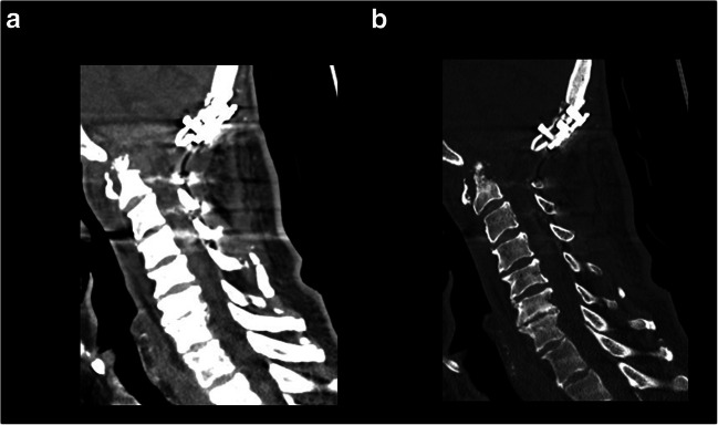
This study aims to describe the posterolateral epidural supra-C2-root approach (PESCA), which might be a good alternative to the transoral, anterolateral, and other posterolateral approaches for biopsy of lesions of the odontoid process (OP). The preoperative planning of PESCA included computerized tomography (CT), CT-angiography, and three-dimensional reconstruction (if possible, even with three-dimensional print) to analyze the angle of the trajectory and the anatomy of the vertebral artery (VA). For PESCA, the patient is positioned under general anesthesia in prone position. In case of an osteolytic lesion with fracture of the OP, an X-ray is performed after positioning to verify anatomic alignment. In the first step, in case of instability and compression of the spinal cord, a craniocervical fusion and decompression is performed (laminectomy of the middle part of the C1 arc and removal of the lower part of the lateral C1 arc). The trajectory is immediately above the C2 root (and under the upper rest of the lateral part of C1 arc). Even if the trajectory is narrowed, it is possible to perform PESCA without relevant traction of the spinal cord. The vertical segment of V3 of the VA at the level of C2 is protected by the vertebral foramen, and the horizontal part of V3 is protected by the remnant upper lateral part of the C1 arc (in case of normal variants). PESCA might be a good choice for biopsy of selected lesions of the OP in same sitting procedure after craniocervical stabilization and decompression.
本研究旨在描述经 C2 上椎旁硬膜外 supra-C2-根入路(PESCA),对于齿状突(OP)病变的活检,它可能是经口前路和其他经后路入路的良好替代方法。PESCA 的术前规划包括计算机断层扫描(CT)、CT 血管造影和三维重建(如有可能,甚至进行三维打印),以分析轨迹角度和椎动脉(VA)的解剖结构。对于 PESCA,患者在全身麻醉下采取俯卧位。对于伴有 OP 骨折的溶骨性病变,在定位后进行 X 线检查以验证解剖对齐。在第一步中,对于不稳定和脊髓受压的情况,进行颅颈融合和减压(C1 弧中部的椎板切除术和外侧 C1 弧下部的切除)。轨迹直接位于 C2 根上方(在 C1 弧外侧部分的上休息处下方)。即使轨迹变窄,也可以在不相关的脊髓牵引下进行 PESCA。VA 的 V3 垂直段在 C2 水平处受椎孔保护,而 V3 的水平部分受 C1 弧外侧残留上部的保护(在正常变异的情况下)。在颅颈稳定和减压后,对于某些 OP 病变,PESCA 可能是在同一次就诊中进行活检的良好选择。