• 文献检索
  • 文档翻译
  • 深度研究
  • 学术资讯
  • Suppr Zotero 插件Zotero 插件
  • 邀请有礼
  • 套餐&价格
  • 历史记录
应用&插件
Suppr Zotero 插件Zotero 插件浏览器插件Mac 客户端Windows 客户端微信小程序
定价
高级版会员购买积分包购买API积分包
服务
文献检索文档翻译深度研究API 文档MCP 服务
关于我们
关于 Suppr公司介绍联系我们用户协议隐私条款
关注我们

Suppr 超能文献

核心技术专利:CN118964589B侵权必究
粤ICP备2023148730 号-1Suppr @ 2026

文献检索

告别复杂PubMed语法,用中文像聊天一样搜索,搜遍4000万医学文献。AI智能推荐,让科研检索更轻松。

立即免费搜索

文件翻译

保留排版,准确专业,支持PDF/Word/PPT等文件格式,支持 12+语言互译。

免费翻译文档

深度研究

AI帮你快速写综述,25分钟生成高质量综述,智能提取关键信息,辅助科研写作。

立即免费体验

超顺磁性氧化铁颗粒(VSOPs)显示出遗传毒性作用,但对人脂肪组织来源的基质细胞(ASCs)无功能影响。

Superparamagnetic Iron Oxide Particles (VSOPs) Show Genotoxic Effects but No Functional Impact on Human Adipose Tissue-Derived Stromal Cells (ASCs).

作者信息

Radeloff Katrin, Ramos Tirado Mario, Haddad Daniel, Breuer Kathrin, Müller Jana, Hochmuth Sabine, Hackenberg Stephan, Scherzad Agmal, Kleinsasser Norbert, Radeloff Andreas

机构信息

Department of Otorhinolaryngology, Head and Neck Surgery, University of Oldenburg, 26122 Oldenburg, Germany.

Department of Otorhinolaryngology, Plastic, Aesthetic and Reconstructive Head and Neck Surgery, University of Wuerzburg, 97080 Wuerzburg, Germany.

出版信息

Materials (Basel). 2021 Jan 7;14(2):263. doi: 10.3390/ma14020263.

DOI:10.3390/ma14020263
PMID:33430323
原文链接:https://pmc.ncbi.nlm.nih.gov/articles/PMC7825809/
Abstract

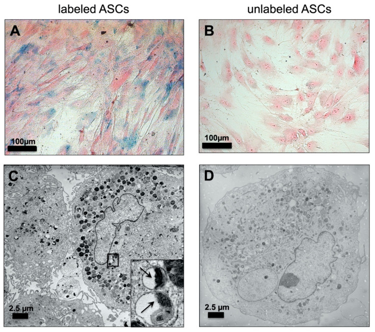
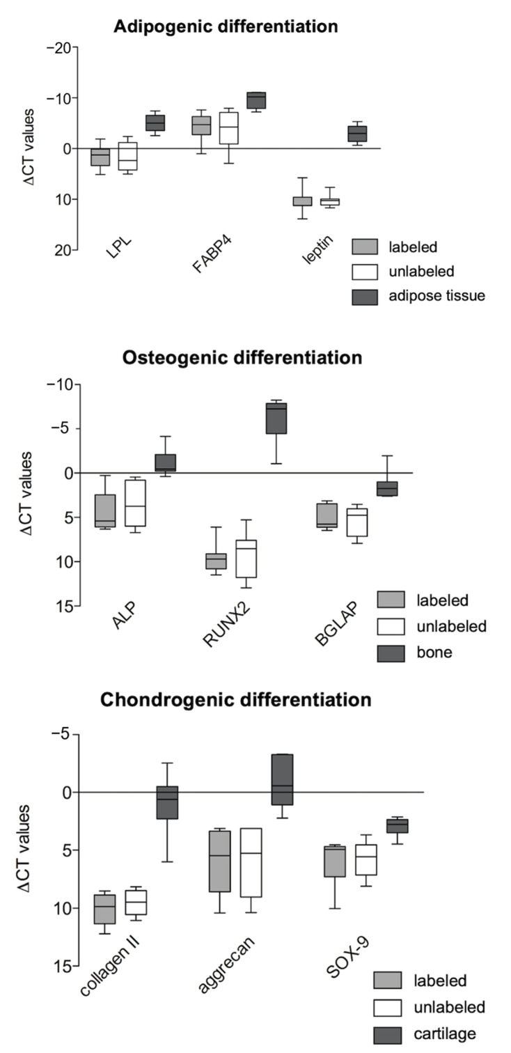
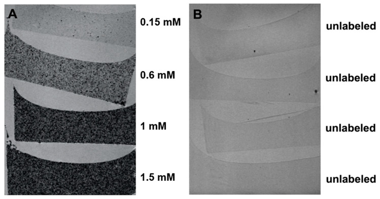
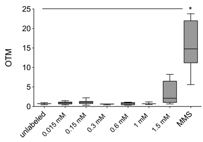
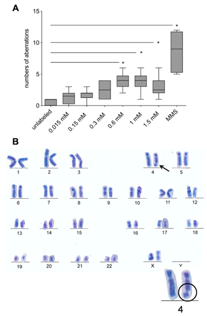
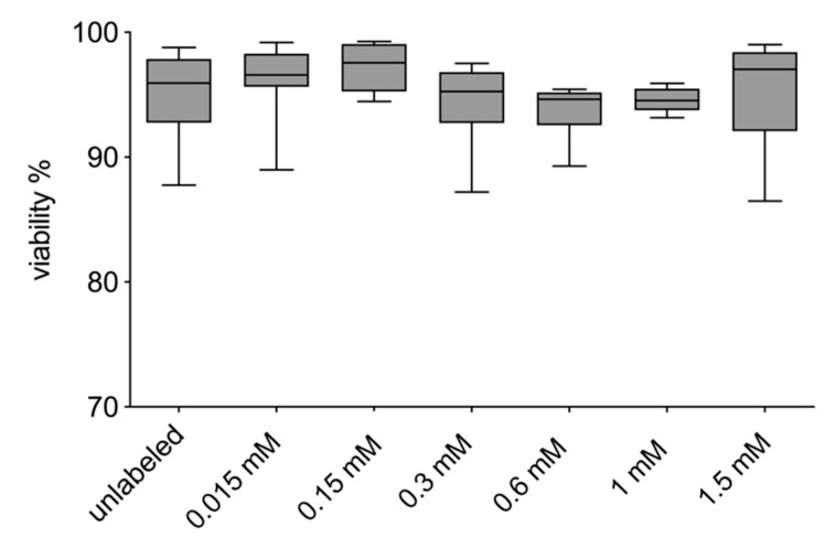
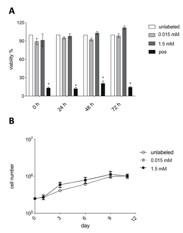
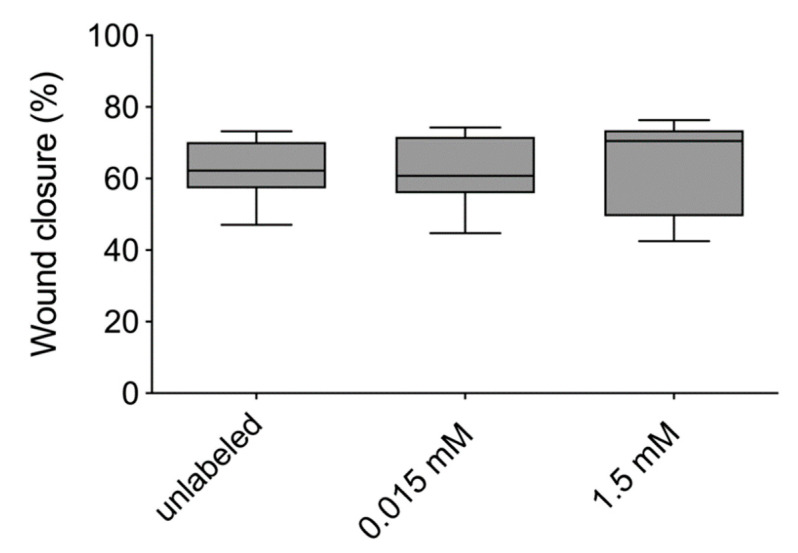
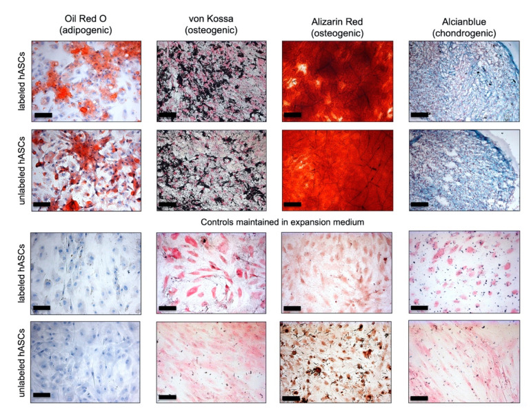
Adipose tissue-derived stromal cells (ASCs) represent a capable source for cell-based therapeutic approaches. For monitoring a cell-based application in vivo, magnetic resonance imaging (MRI) of cells labeled with iron oxide particles is a common method. It is the aim of the present study to analyze potential DNA damage, cytotoxicity and impairment of functional properties of human (h)ASCs after labeling with citrate-coated very small superparamagnetic iron oxide particles (VSOPs). Cytotoxic as well as genotoxic effects of the labeling procedure were measured in labeled and unlabeled hASCs using the MTT assay, comet assay and chromosomal aberration test. Trilineage differentiation was performed to evaluate an impairment of the differentiation potential due to the particles. Proliferation as well as migration capability were analyzed after the labeling procedure. Furthermore, the labeling of the hASCs was confirmed by Prussian blue staining, transmission electron microscopy (TEM) and high-resolution MRI. Below the concentration of 0.6 mM, which was used for the procedure, no evidence of genotoxic effects was found. At 0.6 mM, 1 mM as well as 1.5 mM, an increase in the number of chromosomal aberrations was determined. Cytotoxic effects were not observed at any concentration. Proliferation, migration capability and differentiation potential were also not affected by the procedure. Labeling with VSOPs is a useful labeling method for hASCs that does not affect their proliferation, migration and differentiation potential. Despite the absence of cytotoxicity, however, indications of genotoxic effects have been demonstrated.

摘要

脂肪组织来源的基质细胞(ASCs)是基于细胞的治疗方法的一个可行来源。为了监测体内基于细胞的应用,对用氧化铁颗粒标记的细胞进行磁共振成像(MRI)是一种常用方法。本研究的目的是分析用柠檬酸盐包被的超小超顺磁性氧化铁颗粒(VSOPs)标记后人(h)ASCs的潜在DNA损伤、细胞毒性和功能特性损害。使用MTT法、彗星试验和染色体畸变试验在标记和未标记的hASCs中测量标记过程的细胞毒性和遗传毒性作用。进行三系分化以评估颗粒对分化潜能的损害。在标记过程后分析增殖和迁移能力。此外,通过普鲁士蓝染色、透射电子显微镜(TEM)和高分辨率MRI确认hASCs的标记。在用于该过程的0.6 mM浓度以下,未发现遗传毒性作用的证据。在0.6 mM、1 mM和1.5 mM时,确定染色体畸变数量增加。在任何浓度下均未观察到细胞毒性作用。增殖、迁移能力和分化潜能也不受该过程的影响。用VSOPs标记是一种对hASCs有用的标记方法,不会影响其增殖、迁移和分化潜能。然而,尽管没有细胞毒性,但已证明存在遗传毒性作用的迹象。

https://cdn.ncbi.nlm.nih.gov/pmc/blobs/30cd/7825809/68d0e5f924c0/materials-14-00263-g009.jpg
https://cdn.ncbi.nlm.nih.gov/pmc/blobs/30cd/7825809/1af5bc9cd91f/materials-14-00263-g001.jpg
https://cdn.ncbi.nlm.nih.gov/pmc/blobs/30cd/7825809/da073ad18690/materials-14-00263-g002.jpg
https://cdn.ncbi.nlm.nih.gov/pmc/blobs/30cd/7825809/5f846c8e4957/materials-14-00263-g003.jpg
https://cdn.ncbi.nlm.nih.gov/pmc/blobs/30cd/7825809/d65bbb83d0c4/materials-14-00263-g004.jpg
https://cdn.ncbi.nlm.nih.gov/pmc/blobs/30cd/7825809/ec939fbdc7d7/materials-14-00263-g005.jpg
https://cdn.ncbi.nlm.nih.gov/pmc/blobs/30cd/7825809/d6542c816718/materials-14-00263-g006.jpg
https://cdn.ncbi.nlm.nih.gov/pmc/blobs/30cd/7825809/74dd6b66d730/materials-14-00263-g007.jpg
https://cdn.ncbi.nlm.nih.gov/pmc/blobs/30cd/7825809/031306bbbb0b/materials-14-00263-g008.jpg
https://cdn.ncbi.nlm.nih.gov/pmc/blobs/30cd/7825809/68d0e5f924c0/materials-14-00263-g009.jpg

相似文献

1
Superparamagnetic Iron Oxide Particles (VSOPs) Show Genotoxic Effects but No Functional Impact on Human Adipose Tissue-Derived Stromal Cells (ASCs).超顺磁性氧化铁颗粒(VSOPs)显示出遗传毒性作用,但对人脂肪组织来源的基质细胞(ASCs)无功能影响。
Materials (Basel). 2021 Jan 7;14(2):263. doi: 10.3390/ma14020263.
2
Toxicity and Functional Impairment in Human Adipose Tissue-Derived Stromal Cells (hASCs) Following Long-Term Exposure to Very Small Iron Oxide Particles (VSOPs).长期暴露于超小氧化铁颗粒(VSOPs)后人脂肪组织来源的间充质干细胞(hASCs)的毒性和功能损伤
Nanomaterials (Basel). 2020 Apr 13;10(4):741. doi: 10.3390/nano10040741.
3
DiI labeling of human adipose-derived stem cells: evaluation of DNA damage, toxicity and functional impairment.DiI 标记人脂肪来源干细胞:对 DNA 损伤、毒性和功能障碍的评估。
Cells Tissues Organs. 2013;197(5):384-98. doi: 10.1159/000346714. Epub 2013 Mar 9.
4
Synthesis of acid-stabilized iron oxide nanoparticles and comparison for targeting atherosclerotic plaques: evaluation by MRI, quantitative MPS, and TEM alternative to ambiguous Prussian blue iron staining.酸稳定氧化铁纳米颗粒的合成及靶向动脉粥样硬化斑块的比较:通过磁共振成像、定量多谱勒血管成像和透射电子显微镜评估替代模糊的普鲁士蓝铁染色法
Nanomedicine. 2015 Jul;11(5):1085-95. doi: 10.1016/j.nano.2015.01.002. Epub 2015 Feb 4.
5
Magnetic resonance imaging of ultrasmall superparamagnetic iron oxide-labeled exosomes from stem cells: a new method to obtain labeled exosomes.干细胞来源的超小型超顺磁性氧化铁标记外泌体的磁共振成像:一种获取标记外泌体的新方法。
Int J Nanomedicine. 2016 Jun 1;11:2481-90. doi: 10.2147/IJN.S104152. eCollection 2016.
6
Behaviour of adipose-derived canine mesenchymal stem cells after superparamagnetic iron oxide nanoparticles labelling for magnetic resonance imaging.超顺磁性氧化铁纳米颗粒标记后用于磁共振成像的犬脂肪来源间充质干细胞的行为
BMC Vet Res. 2017 Feb 24;13(1):62. doi: 10.1186/s12917-017-0980-0.
7
Iron oxide particles for molecular magnetic resonance imaging cause transient oxidative stress in rat macrophages.用于分子磁共振成像的氧化铁颗粒会在大鼠巨噬细胞中引发短暂的氧化应激。
Free Radic Biol Med. 2004 Apr 15;36(8):976-84. doi: 10.1016/j.freeradbiomed.2004.01.016.
8
Superparamagnetic iron oxide does not affect the viability and function of adipose-derived stem cells, and superparamagnetic iron oxide-enhanced magnetic resonance imaging identifies viable cells.超顺磁性氧化铁不影响脂肪来源干细胞的活力和功能,且超顺磁性氧化铁增强磁共振成像可识别活细胞。
Magn Reson Imaging. 2009 Jan;27(1):108-19. doi: 10.1016/j.mri.2008.05.013. Epub 2008 Jul 26.
9
Repeated Injection of Very Small Superparamagnetic Iron Oxide Particles (VSOPs) in Murine Atherosclerosis: A Safety Study.在小鼠动脉粥样硬化模型中重复注射超小超顺磁性氧化铁颗粒(VSOPs):一项安全性研究。
Nanomaterials (Basel). 2024 Apr 28;14(9):773. doi: 10.3390/nano14090773.
10
Comparative Labeling of Equine and Ovine Multipotent Stromal Cells With Superparamagnetic Iron Oxide Particles for Magnetic Resonance Imaging In Vitro.用于体外磁共振成像的超顺磁性氧化铁颗粒对马和羊多能基质细胞的对比标记
Cell Transplant. 2015;24(6):1111-25. doi: 10.3727/096368913X675737. Epub 2013 Dec 10.

引用本文的文献

1
Influence of Physicochemical Properties of Iron Oxide Nanoparticles on Their Antibacterial Activity.氧化铁纳米颗粒的物理化学性质对其抗菌活性的影响
ACS Omega. 2024 Jul 25;9(31):33303-33334. doi: 10.1021/acsomega.4c02822. eCollection 2024 Aug 6.
2
Evaluation of In Vitro Cytotoxic, Genotoxic, Apoptotic, and Cell Cycle Arrest Potential of Iron-Nickel Alloy Nanoparticles.铁镍合金纳米颗粒的体外细胞毒性、遗传毒性、凋亡及细胞周期阻滞潜力评估
Toxics. 2022 Aug 24;10(9):492. doi: 10.3390/toxics10090492.
3
Magnetic Nanoparticles Used in Oncology.

本文引用的文献

1
Toxicity and Functional Impairment in Human Adipose Tissue-Derived Stromal Cells (hASCs) Following Long-Term Exposure to Very Small Iron Oxide Particles (VSOPs).长期暴露于超小氧化铁颗粒(VSOPs)后人脂肪组织来源的间充质干细胞(hASCs)的毒性和功能损伤
Nanomaterials (Basel). 2020 Apr 13;10(4):741. doi: 10.3390/nano10040741.
2
Fat Graft Enrichment Strategies: A Systematic Review.脂肪移植富集策略:系统评价。
Plast Reconstr Surg. 2020 Mar;145(3):827-841. doi: 10.1097/PRS.0000000000006557.
3
Biodistribution, Clearance And Morphological Alterations Of Intravenously Administered Iron Oxide Nanoparticles In Male Wistar Rats.
用于肿瘤学的磁性纳米颗粒。
Materials (Basel). 2021 Oct 10;14(20):5948. doi: 10.3390/ma14205948.
4
Differentiation Behaviour of Adipose-Derived Stromal Cells (ASCs) Seeded on Polyurethane-Fibrin Scaffolds In Vitro and In Vivo.接种于聚氨酯-纤维蛋白支架上的脂肪来源基质细胞(ASCs)在体内外的分化行为
Biomedicines. 2021 Aug 9;9(8):982. doi: 10.3390/biomedicines9080982.
静脉注射氧化铁纳米颗粒在雄性 Wistar 大鼠体内的分布、清除和形态学改变。
Int J Nanomedicine. 2019 Dec 6;14:9677-9692. doi: 10.2147/IJN.S223142. eCollection 2019.
4
A Safe and Effective Magnetic Labeling Protocol for MRI-Based Tracking of Human Adult Neural Stem Cells.一种用于基于MRI追踪成人人类神经干细胞的安全有效的磁性标记方案。
Front Neurosci. 2019 Oct 11;13:1092. doi: 10.3389/fnins.2019.01092. eCollection 2019.
5
Targeted magnetic nanoparticle hyperthermia for the treatment of oral cancer.靶向磁性纳米粒子热疗治疗口腔癌。
J Oral Pathol Med. 2019 Oct;48(9):803-809. doi: 10.1111/jop.12921. Epub 2019 Aug 3.
6
Harnessing the secretome of adipose-derived stem cells in the treatment of ischemic heart diseases.利用脂肪来源干细胞的 secretome 治疗缺血性心脏病。
Stem Cell Res Ther. 2019 Jun 27;10(1):196. doi: 10.1186/s13287-019-1289-7.
7
Molecular Mechanisms Responsible for Therapeutic Potential of Mesenchymal Stem Cell-Derived Secretome.间充质干细胞分泌组的治疗潜力的分子机制。
Cells. 2019 May 16;8(5):467. doi: 10.3390/cells8050467.
8
Evaluation of DNA interaction, genotoxicity and oxidative stress induced by iron oxide nanoparticles both in vitro and in vivo: attenuation by thymoquinone.评价氧化铁纳米颗粒在体内外的 DNA 相互作用、遗传毒性和氧化应激:由百里醌进行的衰减。
Sci Rep. 2019 May 6;9(1):6912. doi: 10.1038/s41598-019-43188-5.
9
The mesenchymal stem cell secretome: A new paradigm towards cell-free therapeutic mode in regenerative medicine.间质干细胞分泌组:再生医学中无细胞治疗模式的新范例。
Cytokine Growth Factor Rev. 2019 Apr;46:1-9. doi: 10.1016/j.cytogfr.2019.04.002. Epub 2019 Apr 2.
10
Adipose-derived stem cells: Sources, potency, and implications for regenerative therapies.脂肪来源干细胞:来源、多能性及其在再生治疗中的意义。
Biomed Pharmacother. 2019 Jun;114:108765. doi: 10.1016/j.biopha.2019.108765. Epub 2019 Mar 25.