• 文献检索
  • 文档翻译
  • 深度研究
  • 学术资讯
  • Suppr Zotero 插件Zotero 插件
  • 邀请有礼
  • 套餐&价格
  • 历史记录
应用&插件
Suppr Zotero 插件Zotero 插件浏览器插件Mac 客户端Windows 客户端微信小程序
定价
高级版会员购买积分包购买API积分包
服务
文献检索文档翻译深度研究API 文档MCP 服务
关于我们
关于 Suppr公司介绍联系我们用户协议隐私条款
关注我们

Suppr 超能文献

核心技术专利:CN118964589B侵权必究
粤ICP备2023148730 号-1Suppr @ 2026

文献检索

告别复杂PubMed语法,用中文像聊天一样搜索,搜遍4000万医学文献。AI智能推荐,让科研检索更轻松。

立即免费搜索

文件翻译

保留排版,准确专业,支持PDF/Word/PPT等文件格式,支持 12+语言互译。

免费翻译文档

深度研究

AI帮你快速写综述,25分钟生成高质量综述,智能提取关键信息,辅助科研写作。

立即免费体验

分支型视网膜静脉阻塞中非灌注区分布及严重程度与细胞因子水平和黄斑厚度的关系。

Relationship between distribution and severity of non-perfusion and cytokine levels and macular thickness in branch retinal vein occlusion.

机构信息

Department of Ophthalmology, Yeungnam University College of Medicine, #170 Hyunchungro, Nam-gu, Daegu, 42415, South Korea.

Yeungnam Eye Center, Yeungnam University Hospital, Daegu, South Korea.

出版信息

Sci Rep. 2021 Jan 11;11(1):271. doi: 10.1038/s41598-020-79522-5.

DOI:10.1038/s41598-020-79522-5
PMID:33432033
原文链接:https://pmc.ncbi.nlm.nih.gov/articles/PMC7801443/
Abstract

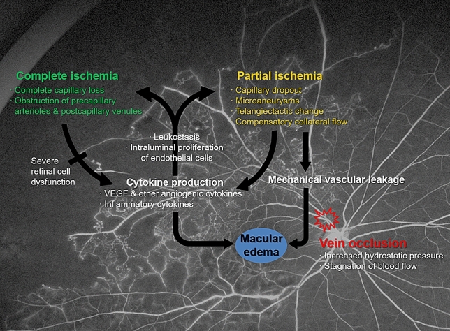
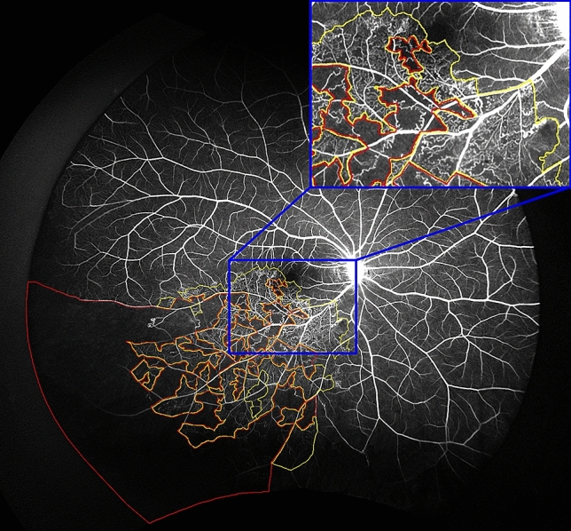
We aimed to investigate the relationship between non-perfusion on ultra-widefield angiography (UWF FA) and aqueous cytokine levels and central macular thickness (CMT) in eyes with branch retinal vein occlusion (BRVO). Thirty-five eyes with treatment-naïve BRVO were included. Non-perfusion area (NPA) for partial and complete ischemia was manually segmented and the ischemic index (ISI) for each was calculated using stereographically projected UWF FA for four different retinal zones. Partial and complete ischemia had different regional predominance. Partial ischemia was predominant in the posterior regions, while complete ischemia was predominant in the periphery. And partial ischemic area, located posterior to far periphery, showed significant correlation with central macular thickness and concentrations of angiogenic and inflammatory cytokines, while complete ischemic area showed no correlation with any of the parameters. Taken together, partial but not complete ischemia, particularly in the more posterior retina, was associated with higher cytokine levels and more severe macular edema in eyes with BRVO. These findings would help us to better understand the different clinical significance of ischemia in BRVO depending on the severity and regional distribution.

摘要

我们旨在研究在未经治疗的视网膜分支静脉阻塞(BRVO)患者中,广角荧光血管造影(UWF FA)无灌注区与房水细胞因子水平和中心黄斑厚度(CMT)之间的关系。共纳入 35 只接受治疗的 BRVO 眼。使用立体投影 UWF FA 对四个不同视网膜区域手动分割部分和完全缺血的无灌注区(NPA),并计算每个区域的缺血指数(ISI)。部分和完全缺血具有不同的区域性优势。部分缺血主要位于后部区域,而完全缺血主要位于周边部。并且位于远周边部后部的部分缺血区与中央黄斑厚度和血管生成及炎症细胞因子浓度有显著相关性,而完全缺血区与任何参数均无相关性。综上所述,在 BRVO 眼中,部分而非完全缺血,特别是在后部更明显的缺血,与更高的细胞因子水平和更严重的黄斑水肿有关。这些发现将帮助我们更好地理解 BRVO 中缺血的不同临床意义,取决于其严重程度和区域分布。

https://cdn.ncbi.nlm.nih.gov/pmc/blobs/9613/7801443/a9694617f50d/41598_2020_79522_Fig2_HTML.jpg
https://cdn.ncbi.nlm.nih.gov/pmc/blobs/9613/7801443/151b4c5b15eb/41598_2020_79522_Fig1_HTML.jpg
https://cdn.ncbi.nlm.nih.gov/pmc/blobs/9613/7801443/a9694617f50d/41598_2020_79522_Fig2_HTML.jpg
https://cdn.ncbi.nlm.nih.gov/pmc/blobs/9613/7801443/151b4c5b15eb/41598_2020_79522_Fig1_HTML.jpg
https://cdn.ncbi.nlm.nih.gov/pmc/blobs/9613/7801443/a9694617f50d/41598_2020_79522_Fig2_HTML.jpg

相似文献

1
Relationship between distribution and severity of non-perfusion and cytokine levels and macular thickness in branch retinal vein occlusion.分支型视网膜静脉阻塞中非灌注区分布及严重程度与细胞因子水平和黄斑厚度的关系。
Sci Rep. 2021 Jan 11;11(1):271. doi: 10.1038/s41598-020-79522-5.
2
Macular Microvascular Changes and Their Correlation With Peripheral Nonperfusion in Branch Retinal Vein Occlusion.黄斑微血管改变及其与分支型视网膜静脉阻塞外周无灌注的相关性。
Am J Ophthalmol. 2021 May;225:57-68. doi: 10.1016/j.ajo.2020.12.026. Epub 2021 Jan 4.
3
Aqueous Angiopoietin-Like 4 Levels Correlate With Nonperfusion Area and Macular Edema in Branch Retinal Vein Occlusion.房水血管生成素样4水平与视网膜分支静脉阻塞中的无灌注区和黄斑水肿相关。
Invest Ophthalmol Vis Sci. 2016 Jan 1;57(1):6-11. doi: 10.1167/iovs.15-18304.
4
Changes in retinal ischaemic index correlate with recalcitrant macular oedema in retinal vein occlusion: WAVE study.视网膜缺血指数的变化与视网膜静脉阻塞性顽固性黄斑水肿相关:WAVE 研究。
Br J Ophthalmol. 2018 Aug;102(8):1066-1071. doi: 10.1136/bjophthalmol-2017-311475. Epub 2018 Apr 26.
5
Distribution of leakage index using ultra-widefield fluorescein angiography in patients with non-ischemic branch retinal vein occlusion and its association with macular edema.利用超广角荧光素血管造影术评估非缺血型分支视网膜静脉阻塞患者的渗漏指数及其与黄斑水肿的关系。
Photodiagnosis Photodyn Ther. 2023 Sep;43:103731. doi: 10.1016/j.pdpdt.2023.103731. Epub 2023 Aug 5.
6
Comparison of aqueous concentrations of angiogenic and inflammatory cytokines in diabetic macular oedema and macular oedema due to branch retinal vein occlusion.糖尿病性黄斑水肿和分支视网膜静脉阻塞所致黄斑水肿中血管生成和炎症细胞因子的水浓度比较。
Br J Ophthalmol. 2012 Nov;96(11):1426-30. doi: 10.1136/bjophthalmol-2012-301913. Epub 2012 Aug 28.
7
Changes in aqueous cytokines after intravitreal triamcinolone versus bevacizumab for macular oedema in branch retinal vein occlusion.玻璃体内注射曲安奈德与贝伐单抗治疗视网膜分支静脉阻塞性黄斑水肿后房水细胞因子的变化
Acta Ophthalmol. 2014 May;92(3):e217-24. doi: 10.1111/aos.12219. Epub 2013 Jul 26.
8
Intravitreal bevacizumab and cytokine levels in major and macular branch retinal vein occlusion.玻璃体内贝伐单抗与大分支和黄斑分支视网膜静脉阻塞的细胞因子水平。
Ophthalmologica. 2011;225(3):150-4. doi: 10.1159/000322364. Epub 2010 Dec 9.
9
Association of aqueous humor cytokines with the development of retinal ischemia and recurrent macular edema in retinal vein occlusion.房水细胞因子与视网膜静脉阻塞中视网膜缺血和复发性黄斑水肿发展的关系。
Invest Ophthalmol Vis Sci. 2014 Apr 9;55(4):2290-6. doi: 10.1167/iovs.13-13587.
10
Aqueous humor inflammatory cytokine levels and choroidal thickness in patients with macular edema associated with branch retinal vein occlusion.伴有分支视网膜静脉阻塞的黄斑水肿患者房水炎性细胞因子水平与脉络膜厚度。
Int Ophthalmol. 2021 Jul;41(7):2433-2444. doi: 10.1007/s10792-021-01798-x. Epub 2021 Mar 19.

引用本文的文献

1
Subretinal fluid in macular edema secondary to branch retinal vein occlusion.视网膜分支静脉阻塞继发黄斑水肿中的视网膜下液
Sci Rep. 2024 Jun 13;14(1):13623. doi: 10.1038/s41598-024-64047-y.
2
Relationship between ischemic index, leakage index, and macular edema in branch retinal vein occlusion.分支型视网膜静脉阻塞中缺血指数、渗漏指数与黄斑水肿的关系。
Graefes Arch Clin Exp Ophthalmol. 2024 May;262(5):1499-1506. doi: 10.1007/s00417-023-06343-3. Epub 2023 Dec 26.
3
Clinical utility of ultra-widefield fluorescein angiography and optical coherence tomography angiography for retinal vein occlusions.

本文引用的文献

1
Cytokines and the Pathogenesis of Macular Edema in Branch Retinal Vein Occlusion.细胞因子与视网膜分支静脉阻塞性黄斑水肿的发病机制
J Ophthalmol. 2019 May 2;2019:5185128. doi: 10.1155/2019/5185128. eCollection 2019.
2
Ultra-Wide-Field Fluorescein Angiography-Guided Normalization of Ischemic Index Calculation in Eyes With Retinal Vein Occlusion.超广域荧光素血管造影引导视网膜静脉阻塞患眼缺血指数计算的标准化。
Invest Ophthalmol Vis Sci. 2018 Jul 2;59(8):3278-3285. doi: 10.1167/iovs.18-23796.
3
Changes in retinal ischaemic index correlate with recalcitrant macular oedema in retinal vein occlusion: WAVE study.
超广角荧光素血管造影和光学相干断层扫描血管造影在视网膜静脉阻塞中的临床应用
Front Med (Lausanne). 2023 Jun 8;10:1110166. doi: 10.3389/fmed.2023.1110166. eCollection 2023.
4
Non-perfusion severity correlates with central macular thickness and microvascular impairment in branch retinal vein occlusions.非灌注严重程度与分支视网膜静脉阻塞的中心黄斑厚度和微血管损伤相关。
Eur J Ophthalmol. 2024 Jan;34(1):226-232. doi: 10.1177/11206721231175525. Epub 2023 May 11.
5
Deep Capillary Plexus as Biomarker of Peripheral Capillary Nonperfusion in Central Retinal Vein Occlusion.深层毛细血管丛作为视网膜中央静脉阻塞中周边毛细血管无灌注的生物标志物
Ophthalmol Sci. 2023 Jan 2;3(2):100267. doi: 10.1016/j.xops.2022.100267. eCollection 2023 Jun.
6
Analysis of the Cytokine Expression in the Aqueous Humor of Individuals with BRVO-Associated Macular Edema.视网膜分支静脉阻塞相关性黄斑水肿患者房水中细胞因子表达分析
J Ophthalmol. 2022 Aug 4;2022:1514244. doi: 10.1155/2022/1514244. eCollection 2022.
7
Review: The Development of Risk Factors and Cytokines in Retinal Vein Occlusion.综述:视网膜静脉阻塞中危险因素及细胞因子的发展
Front Med (Lausanne). 2022 Jun 15;9:910600. doi: 10.3389/fmed.2022.910600. eCollection 2022.
8
Efficacy of intravitreal conbercept injection on short- and long-term macular edema in branch retinal vein occlusion.玻璃体内注射康柏西普治疗视网膜分支静脉阻塞所致短期及长期黄斑水肿的疗效
Int J Ophthalmol. 2022 Mar 18;15(3):489-494. doi: 10.18240/ijo.2022.03.18. eCollection 2022.
视网膜缺血指数的变化与视网膜静脉阻塞性顽固性黄斑水肿相关:WAVE 研究。
Br J Ophthalmol. 2018 Aug;102(8):1066-1071. doi: 10.1136/bjophthalmol-2017-311475. Epub 2018 Apr 26.
4
Distribution of Nonperfusion Area on Ultra-widefield Fluorescein Angiography in Eyes With Diabetic Macular Edema: DAVE Study.糖尿病性黄斑水肿患者超广角荧光素血管造影中非灌注区的分布:DAVE研究
Am J Ophthalmol. 2017 Aug;180:110-116. doi: 10.1016/j.ajo.2017.05.024. Epub 2017 Jun 1.
5
Peripheral Laser for Recalcitrant Macular Edema Owing to Retinal Vein Occlusion: The WAVE Trial.视网膜静脉阻塞所致顽固性黄斑水肿的周边激光治疗:WAVE试验
Ophthalmology. 2017 Jun;124(6):919-921. doi: 10.1016/j.ophtha.2017.01.049. Epub 2017 Mar 18.
6
Measuring the precise area of peripheral retinal non-perfusion using ultra-widefield imaging and its correlation with the ischaemic index.使用超广角成像测量周边视网膜无灌注的确切面积及其与缺血指数的相关性。
Br J Ophthalmol. 2016 Feb;100(2):235-9. doi: 10.1136/bjophthalmol-2015-306652. Epub 2015 Jul 1.
7
Scatter Photocoagulation Does Not Reduce Macular Edema or Treatment Burden in Patients with Retinal Vein Occlusion: The RELATE Trial.散射光凝术不能减轻视网膜静脉阻塞患者的黄斑水肿或治疗负担:RELATE试验
Ophthalmology. 2015 Jul;122(7):1426-37. doi: 10.1016/j.ophtha.2015.04.006. Epub 2015 May 9.
8
Assessment of accuracy and precision of quantification of ultra-widefield images.超广角图像量化的准确性和精密度评估。
Ophthalmology. 2015 Apr;122(4):864-6. doi: 10.1016/j.ophtha.2014.11.016. Epub 2015 Jan 8.
9
Precise montaging and metric quantification of retinal surface area from ultra-widefield fundus photography and fluorescein angiography.通过超广角眼底摄影和荧光素血管造影对视网膜表面积进行精确拼接和测量量化。
Ophthalmic Surg Lasers Imaging Retina. 2014 Jul-Aug;45(4):312-7. doi: 10.3928/23258160-20140709-07.
10
Area of peripheral retinal nonperfusion and treatment response in branch and central retinal vein occlusion.视网膜分支静脉阻塞和视网膜中央静脉阻塞中周边视网膜无灌注区及治疗反应
Retina. 2014 Sep;34(9):1736-42. doi: 10.1097/IAE.0000000000000148.