Cui LiHuang, Xiang ShouYang, Chen DeChun, Fu Rui, Zhang Xin, Chen JingTao, Wang XinTao
Department of Orthopedic Surgery, The Second Affiliated Hospital of Harbin Medical University, Harbin, China.
J Orthop Translat. 2020 Sep 14;26:151-161. doi: 10.1016/j.jot.2020.06.003. eCollection 2021 Jan.
Autogenous bone graft is the gold standard bone grafting substrate available in spinal fusion because of its osteoconductive, osteogenic, and osteoinductive properties. However, several shortcomings including bleeding, infection, chronic pain, and nerve injury are known to be associated with the procedure. Bone tissue engineering has emerged as an alternative therapeutic strategy for bone grafts. New materials have been developed and tested that can substitute for the autogenous bone grafts used in the spinal fusion. The purpose of this study is to evaluate the role of a novel tissue-engineered bone graft with silicon-substituted calcium phosphate (Si-CaP), autogenous fine particulate bone powder (AFPBP), and bone marrow mesenchymal stem cells (BMSCs) using a rabbit posterolateral lumbar fusion model based on bone tissue engineering principles. The application of this graft can represent a novel choice for autogenous bone to reduce the amount of autogenous bone and promote spinal fusion.
BMSCs from New Zealand white rabbits were isolated and cultured in vitro. Then, BMSCs were marked by the cell tracker chloromethyl-benzamidodialkylcarbocyanine (CM-Dil). A total of 96 New Zealand White rabbits were randomly divided into four groups: (a) AFPBP, (b) Si-CaP, (c) Si-CaP/AFPBP, (d) Si-CaP/AFPBP/BMSCs.The rabbits underwent bilateral posterolateral spine arthrodesis of the L5-L6 intertransverse processes using different grafts. Spinal fusion and bone formation were evaluated at 4, 8, and 12 weeks after surgery by manual palpation, radiology, micro-computed tomography (micro-CT), histology, and scanning electronic microscopy (SEM).
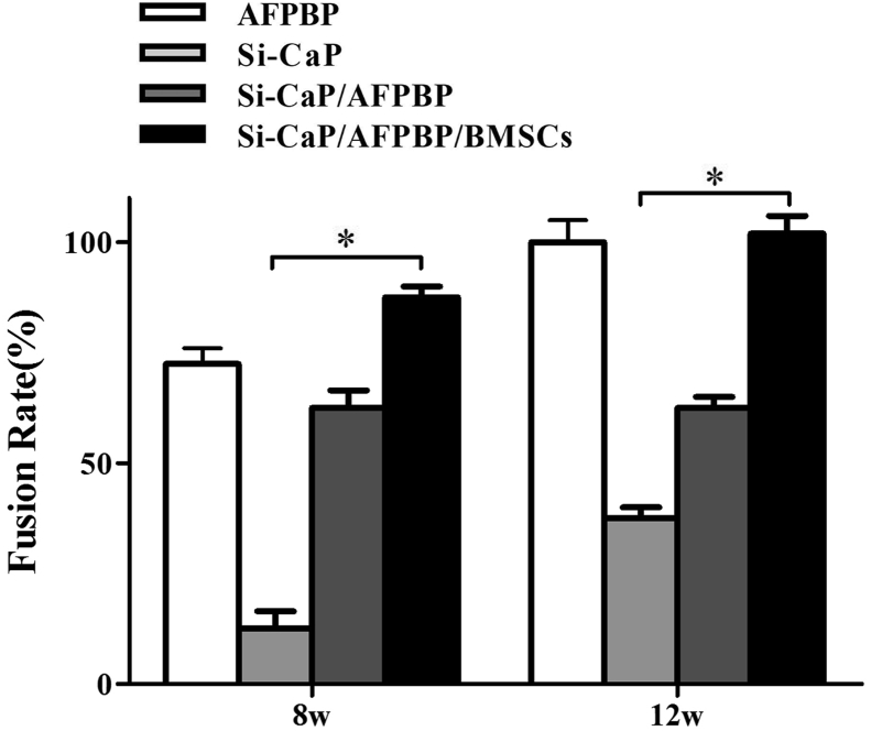
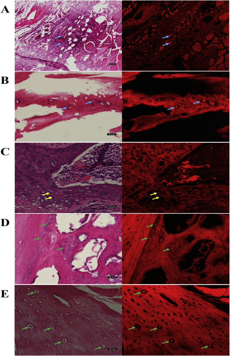
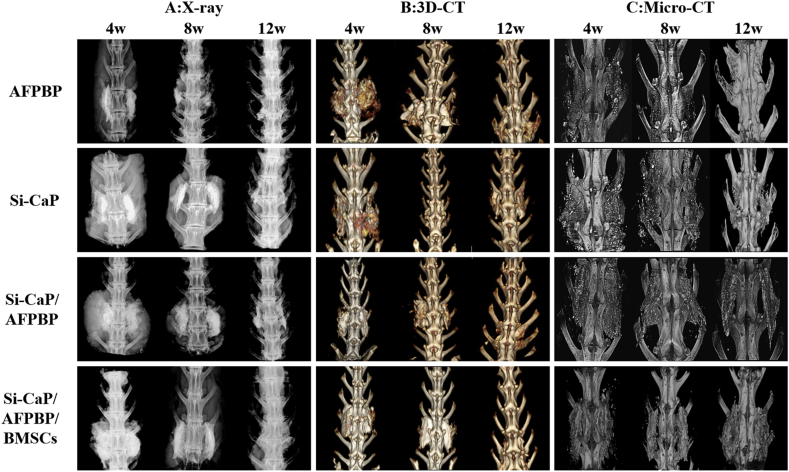
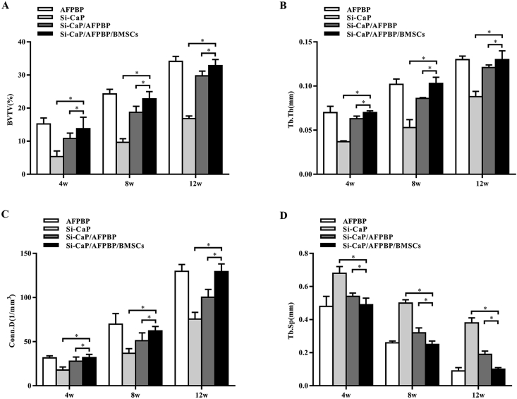
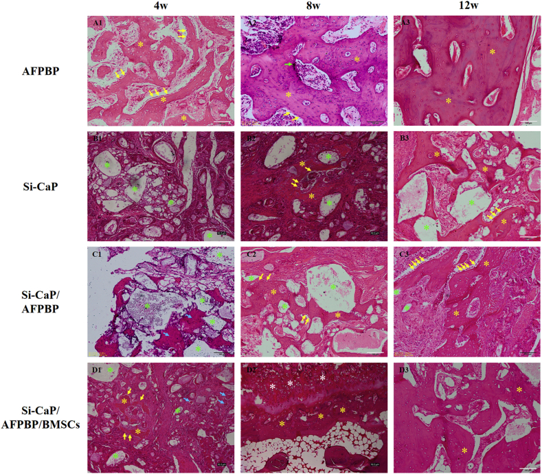
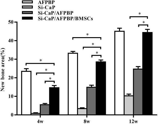
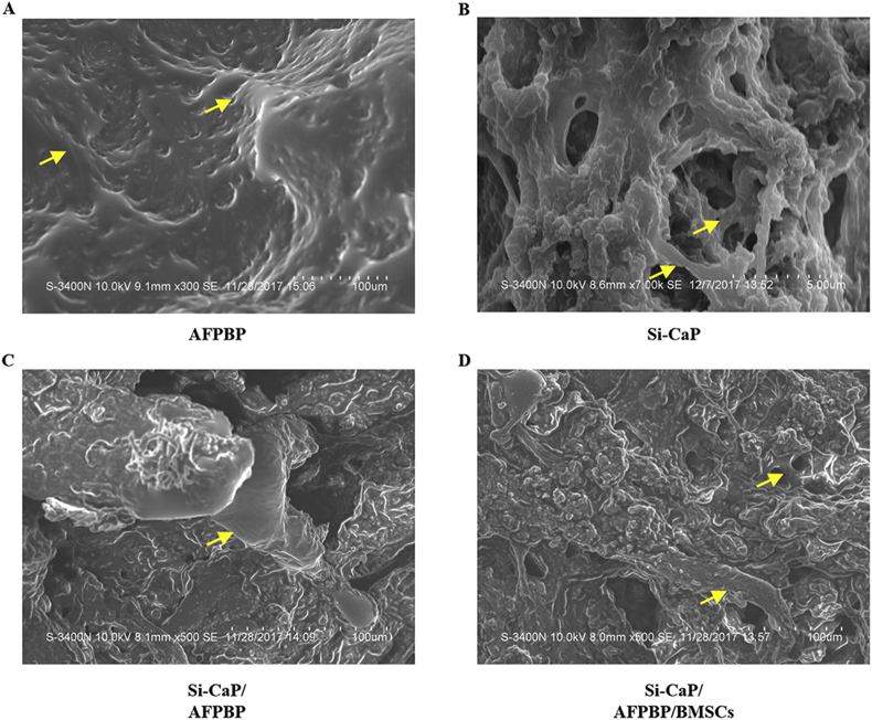
The rate of fusion by manual palpation was higher in the Si-CaP/AFPBP/BMSCs group than the other groups at 8 weeks. The fusion rates in the Si-CaP/AFPBP/BMSCs and the AFPBP groups both reached 100%, which was higher than the Si-CaP/AFPBP group (62.5%) (P > 0.05) and Si-CaP group (37.5%) (P < 0.05) at 12 weeks. New bone formation was observed in all groups after implantation by radiology and micro-CT. The radiographic and CT scores increased in all groups from 4 to 12 weeks, indicating a time-dependent osteogenetic process. The Si-CaP/AFPBP/BMSCs group showed a larger amount of newly formed bone than the Si-CaP/AFPBP and Si-CaP groups at 12 weeks. Bone formation in the Si-CaP/AFPBP/BMSCs group was similar to the AFPBP group. Histology showed that new bone formation continued and increased along with the degradation and absorption of Si-CaP and AFPBP from 4 to 12 weeks in the Si-CaP, Si-CaP/AFPBP, and Si-CaP/AFPBP/BMSCs groups. At 4 weeks, a higher proportion of bone was detected in the AFPBP group (23.49%) compared with the Si-CaP/AFPBP/BMSCs group (14.66%, P < 0.05). In the Si-CaP/AFPBP/BMSCs group at 8 weeks, the area percentage of new bone formation was 28.56%, which was less than the AFPBP group (33.21%, P < 0.05). No difference in bone volume was observed between the Si-CaP/AFPBP/BMSCs group (44.39%) and AFPBP group (45.06%) at 12 weeks (P > 0.05). At 12 weeks, new trabecular were visible in the Si-CaP/AFPBP/BMSCs group by SEM. CM-Dil-positive cells were observed at all stages. Compared with histological images, BMSCs participate in various stages of osteogenesis by transforming into osteoblasts, chondrocytes, and osteocytes.
This study demonstrated for the first time that Si-CaP/AFPBP/BMSCs is a novel tissue-engineered bone graft with excellent bioactivity, biocompatibility, and biodegradability. The graft could reduce the amount of autogenous bone and promote spinal fusion in a rabbit posterolateral lumbar fusion model, representing a novel alternative to autogenous bone.
The translational potential of this article lies in that this graft will be a novel spinal fusion graft with great potential for clinical applications.
自体骨移植因其具有骨传导性、成骨性和骨诱导性,是脊柱融合术中可用的金标准骨移植基质。然而,已知该手术存在包括出血、感染、慢性疼痛和神经损伤等若干缺点。骨组织工程已成为骨移植的一种替代治疗策略。已开发并测试了可替代脊柱融合术中使用的自体骨移植的新材料。本研究的目的是基于骨组织工程原理,使用兔腰椎后外侧融合模型评估一种新型组织工程骨移植(含硅取代磷酸钙(Si-CaP)、自体细颗粒骨粉(AFPBP)和骨髓间充质干细胞(BMSCs))的作用。这种移植的应用可为自体骨提供一种新的选择,以减少自体骨的用量并促进脊柱融合。
从新西兰白兔分离骨髓间充质干细胞并进行体外培养。然后,用细胞追踪剂氯甲基苯甲酰二烷基碳菁(CM-Dil)标记骨髓间充质干细胞。将96只新西兰白兔随机分为四组:(a)AFPBP组,(b)Si-CaP组,(c)Si-CaP/AFPBP组,(d)Si-CaP/AFPBP/BMSCs组。使用不同的移植物对兔进行L5-L6横突间双侧后外侧脊柱融合术。术后4周、8周和12周通过手法触诊、放射学、微型计算机断层扫描(micro-CT)、组织学和扫描电子显微镜(SEM)评估脊柱融合和骨形成情况。
在8周时,Si-CaP/AFPBP/BMSCs组通过手法触诊的融合率高于其他组。在12周时,Si-CaP/AFPBP/BMSCs组和AFPBP组的融合率均达到100%,高于Si-CaP/AFPBP组(62.5%)(P>0.05)和Si-CaP组(37.5%)(P<0.05)。放射学和micro-CT检查显示,植入后所有组均观察到新骨形成。所有组的放射学和CT评分从4周增加到12周,表明存在时间依赖性的成骨过程。在12周时,Si-CaP/AFPBP/BMSCs组显示出比Si-CaP/AFPBP组和Si-CaP组更多的新形成骨。Si-CaP/AFPBP/BMSCs组的骨形成与AFPBP组相似。组织学显示,在Si-CaP组、Si-CaP/AFPBP组和Si-CaP/AFPBP/BMSCs组中,从4周到12周,随着Si-CaP和AFPBP的降解和吸收,新骨形成持续并增加。在4周时,AFPBP组检测到的骨比例(23.49%)高于Si-CaP/AFPBP/BMSCs组(14.66%,P<0.05)。在8周时,Si-CaP/AFPBP/BMSCs组新骨形成的面积百分比为28.56%,低于AFPBP组(33.21%,P<0.05)。在12周时,Si-CaP/AFPBP/BMSCs组(44.39%)和AFPBP组(45.06%)之间的骨体积无差异(P>0.05)。在12周时,通过SEM在Si-CaP/AFPBP/BMSCs组可见新的小梁。在所有阶段均观察到CM-Dil阳性细胞。与组织学图像相比,骨髓间充质干细胞通过转化为成骨细胞、软骨细胞和骨细胞参与成骨的各个阶段。
本研究首次证明Si-CaP/AFPBP/BMSCs是一种具有优异生物活性、生物相容性和生物降解性的新型组织工程骨移植。在兔腰椎后外侧融合模型中,该移植物可减少自体骨用量并促进脊柱融合,是自体骨的一种新的替代物。
本文的转化潜力在于这种移植物将是一种具有巨大临床应用潜力的新型脊柱融合移植物。